Category Archives: Science

The use of information theory for the evaluation of biomarkers of aging and physiological age for prediction of increased risk of aging-related diseases and frailty: rationale and methodology

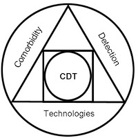
CDT-LOGOBy David Blokh and Ilia Stambler


Summary

This article argues for the expanded application of information-theoretical measures, such as entropy and normalized mutual information, for research of biomarkers of aging and physiological age as an early predictive measure of age-related multimorbidity and frailty. The use of information theory enables unique methodological advantages for the study of aging processes, as it allows to evaluate non-linear relations between biological parameters, providing the precise quantitative strength of those relations, both for individual and multiple parameters, showing cumulative or holistic (synergistic) effects. The diagnostic models can be built based on diagnostic parameters routinely available to physicians (frailty indexes, laboratory analysis, physical evaluations) as well as more advanced biomarkers (e.g. genetic and epigenetic analysis) – in relation with age and age-related diseases and frailty. The diagnostic systems that are built in this way can be open and can include any number of additional parameters correlated with age and age-related diseases. The use of information-theoretical methods, utilizing normalized mutual information, can reveal the exact amount of information that various diagnostic parameters or their combinations contain about the persons’ physiological (or biological) age. Based on those exact diagnostic values for physiological age determination, it is possible to construct a diagnostic decision rule to evaluate a person’s physiological age, as compared to chronological age. The working hypothesis is that people characterized by higher physiological age will have increased risk of age-related frailty and diseases (e.g. heart disease, cancer, type 2 diabetes, neurodegenerative diseases, fractures, falls, mental and functional decline, etc.). Utilizing information-theoretical measures, with additional data, it may be possible to create further clinically applicable information-theory-based markers and models for the evaluation of physiological age, its relation to age-related diseases and its potential modifications by therapeutic interventions, such as medications and behavioral interventions.[1]

Introduction: The increasing need for anti-aging intervention and longevity medicine

With the rapidly growing aging population, and the corresponding rise in the incidence of aging-related diseases (such as heart disease, cancer, type 2 diabetes, neurodegenerative disease, chronic obstructive pulmonary diseases, etc), there emerges a special need to estimate health conditions and effectiveness of treatments for a variety of aging-related diseases, based on the evaluation of the aging processes underlying those diseases. Such evaluation is also needed to assess the effectiveness of potential anti-aging interventions and interventions against aging-related diseases. Even more importantly, it is needed for an early preventive intervention in these diseases, based on the calculated physiological age. The importance of quantifying the effects of “normal” aging as compared to “abnormal”, “pathological”, “accelerated” or “premature” aging cannot be overestimated. It is critically important to be able to diagnose “early aging”, that is, to identify subjects in whom “biological” or “physiological age” markedly exceeds the “chronological age”. Thanks to such “early diagnosis” of aging, as a pre-clinical or concomitant condition for a variety of aging-related diseases, it may be possible to solve the problems of early diagnosis of those aging-derived diseases. In other words, it may be stated that pre-clinical diagnosis of aging-related diseases (such as Alzheimer’s disease, type 2 diabetes, cancer and heart disease) naturally belongs in the field of aging research, as aging can be seen as a pre-symptomatic, pre-clinical root determinant of a variety of aging-related diseases.[2]


Cancer

Of special importance, early evaluation of physiological age may facilitate the early diagnosis of cancer with a prolonged preclinical period. This may considerably improve the efficacy of treatment for oncological diseases. There have been debates regarding the usefulness or lack thereof of mammography for subjects aged 40-49, that is regarding the possibility of preclinical diagnosis.[3] It may be difficult to solve this problem in the framework of pure oncology, disregarding the factors of age or aging, but only in the framework of aging research, for which information-theoretical analysis can be meaningfully applied.


Heart disease

The same may be said for cardiovascular diseases, the main age-related cause of death in the world, including deaths due to ischemic heart disease and ischemic and hemorrhagic stroke.[4] Yet, it is also known that cardiovascular diseases, and ischemic heart disease in particular, can be highly susceptible to therapeutic and lifestyle interventions, capable of dramatically extending the health and longevity of the subjects.[5] Hence it is of primary importance to be able to early assess the entire array of risk factors as well as the effects of therapeutic interventions on the risk factors, either individually or in combinations, including both biological and chronological age.[6]


Neurodegenerative diseases

Also for neurodegenerative diseases, such as Alzheimer’s disease, the vast plasticity of the brain of the aged and the feasibility of positive therapeutic interventions, or even cures, have been recognized. Yet, it has also been recognized that, in order to accomplish such interventions, the earliest possible detection and the consideration of polygenic etiologies will be necessary.[7] Here too information-theoretical statistics, capable of utilizing time series methods for prediction from an earlier, preclinical age, and employing mutual information measures to establish non-linear diagnostic correlations of multiple disease determinants, including age, can be indispensable.


Diabetes – Metabolic syndrome

Also, for type 2 diabetes, “diagnosis of aging” can be very helpful for early diagnosis of diabetes, as the diagnostic parameters relevant to diabetes, as well as the underlying biological mechanisms have a great similarity with “normal” aging.[8]


Frailty

Generally, the ability to reliably quantitatively diagnose “delayed aging” or “healthy aging” may pinpoint powerful factors facilitating healthy and productive longevity. Here “healthy aging” (or “healthy longevity”) may be understood as the absence of age-related frailty, as commonly defined in geriatric medicine, that is, an active and functional state of older adults characterized by a decreased risk for future poor clinical outcomes, diminished development of disability, dementia, falls, hospitalization, institutionalization or decreased mortality.[9] The ability to provide early quantitative evaluation of frailty risks is also of great medical and economic significance.

Prospective economic benefits from the early detection of aging-related diseases thanks to improved diagnosis of aging itself

The humanitarian and economic importance of early detection of aging-related diseases is obvious. Early detection makes it possible to apply preventive medical interventions when the disease is in a more manageable and even curable state, ideally even before any clinical manifestations, thus significantly postponing the time it may take for the disease to progress to a severe, debilitating and more costly state. This postponement of morbidity is also the reason for the vast economic benefits of early detection, necessary for the early preventive intervention. As shown for the US, patients with chronic age-related diseases expend in their last year of life about one third of the total Medicare expenditures (~$15,000 per person).[10] Any postponement of this high morbidity period thanks to early detection and preventive interventions can produce massive net health and economic benefits.

Some of the economic benefits of early detection derive from the improvement of individual health, averting direct medical costs and entitlement payments, reducing lost productivity, disability, and employee turnover.[11] As of 2004, it was estimated that “75 percent of the $1.9 trillion spent on health care in the United States stem from preventable chronic health conditions … but only 1 percent is allocated to protecting health and preventing illness.”[12]

Specifically, regarding particular aging related diseases, such as Alzheimer’s disease and Cancer, the savings from early detection per patient are commonly estimated at several thousand dollars for the developed countries — $1,000-10,000+ for Alzheimer’s disease,[13] $1,000-10,000+ for various forms of cancer.[11] Comparable savings can be expected from the early detection of heart disease[14] and diabetes.[15]

The numbers of patients suffering from these conditions globally are estimated at tens of millions, and are expected to strongly increase worldwide due to the rapid population aging.[16] Thus, 36 million people worldwide are living with dementia, including ~10M in Europe and ~5M in the US, with their numbers expected to double every 20 years, reaching 66 million by 2030, and 115 million by 2050.[13] Out of the total of 56 million deaths that occurred worldwide in 2012, about 38 million were due to non-communicable (NCD) aging-related diseases, in particular: cardiovascular diseases (17.5 million deaths, or 46.2% of NCD deaths), cancers (8.2 million, or 21.7% of NCD deaths), respiratory diseases, including asthma and chronic obstructive pulmonary disease (4.0 million, or 10.7% of NCD deaths) and diabetes (1.5 million, or 4% of NCD deaths).[17]

The costs of aging-related diseases worldwide are correspondingly vast, amounting to hundreds of billions and trillions of dollars: ~US$600 billion in 2010 only for dementia worldwide[13], approximately US$800 billion for heart disease; US$850 billion for type 2 diabetes; US$900 billion for cancer; US$300 billion for Chronic Obstructive Pulmonary Disease – COPD.[18]

Thus the healthcare benefits of even minor improvement in the ability of early diagnosis, necessary for early preventive treatments of aging-related diseases, could be immense. The economic benefits from the preventive approach, intervening in the aging processes underlying the non-communicable diseases before they take clinical forms, would be immense as well (hundreds of billions of dollars savings in health expenditures in the course of several decades just in the US, according to some models [19]). Yet, to accomplish this, improved diagnostic capabilities are needed for the aging process itself, capable to reliably estimate the person’s physiological and biological age and the effects of interventions on that age.


New methodologies are needed to provide early diagnosis of aging-related ill health

In view of the pressing global social need, new methodologies are required to enable early detection of aging-related ill health. We argue that information-theory-based approaches, utilizing such measures as entropy and mutual information, may provide powerful methodological tools for the solution of these problems. First of all, information theory may allow a more reliable estimation of biological and physiological correlates (biomarkers) of aging, due to its ability to estimate non-linear correlations between parameters, utilizing mutual information measures. The a priori reliance on linear statistical correlations when trying to determine such biomarkers has been failing to produce practically applicable results.[20] [21] Information-theoretical measures may provide new means to intensify and facilitate this search. Moreover, the preclinical diagnosis requires the simultaneous analysis of a large number of parameters of various kinds, including continuous parameters, with both Gaussian and non-Gaussian distribution, as well as discrete and ranked parameters. Presently, the only theoretically grounded method for the simultaneous analysis of multiple parameters of different kinds is information theory..[22]


Advantages of the information-theoretical methodology

Arguably, it is methodologically problematic to use the current approaches in biomarkers research and quantified health for practical assessments of physiological age and potential aging-ameliorating and healthspan-extending interventions. The methodological difficulties may derive from two major current shortcomings. Firstly, the current approaches, both in quantified health and biomarkers research, are mainly based on static or short term, average or median population values to define the norm. This makes personalization of clinical evaluations and treatments difficult. Secondly and crucially, the existing approaches commonly assume normal (Gaussian) distribution and linear relations of parameters. Hence, they mainly employ linear statistical measures of correlation, such as the correlation coefficient or linear regression. However, such measures do not correspond to physiological realities, where the relations between parameters are non-linear, including the non-linear alterations with age. Hence the currently used methods are ill suited to evaluate physiological age and aging-ameliorating and healthspan extending interventions. The main advantage of the information-theoretical methodology is that it provides an integrated approach that will take into consideration the non-linear interrelation of a multitude of parameters – biomarkers and intervention factors, using information theoretical measures rather than linear statistical measures.[1]

Information theory can serve as a universal methodology to assess health and disease status, in relation to age, unifying a variety of model systems, focusing on age-related changes as the root cause of a variety of chronic age-related diseases and health impairments. Information theory may provide the following specific methodological capabilities, currently not available in any other system:

  • The current health metrics mainly employ statistical measures. Yet, statistical measures are often inadequate, insofar as in biological systems, the relations between parameters are often non-linear. In contrast, information-theoretical methods allow for the estimation (measurement) of complex non-linear relations between parameters, hence they allow for the inclusion of a wider range of data for making health decisions.
  • Currently, the results from different study models are described in incompatible terms, that do not permit an easy mutual inference. In contrast, the common terms and measures of information theory, such as entropy and mutual information, can serve as a universal language to describe, in a unified way, any number of diverse models and results.
  • Currently, the degree of mutual applicability between animal model systems and humans, as well as between diverse human samples, is uncertain.  In contrast, the evaluation of mutual information between different model systems, can be used as a standardized and convenient estimate of their mutual applicability.
  • Currently, the effects of various treatments on human health are often examined in a disconnected manner, without knowing the precise interactions of various treatments. The information-theoretical measures of correlation (such as normalized mutual information) can be employed to test the effects of single or combinations of various treatment factors (such as drugs, genes and lifestyle factors) on the health span and the disease status. By the precise quantitative evaluation of the influence of such factors on the health span and disease status, both synergistic positive and antagonistic adverse effects of treatment interactions will be determined.
  • The current systems lack the formal ability to select the most informative (and hence clinically useful) parameters. Using information-theoretical methods, the most informative single parameters or groups of parameters with the highest influence on the health span and disease status can be selected. The selection of the most informative parameters, such as those that contain information about other selected parameters, will allow for a more economic, convenient and efficient diagnostic system. This will save time and expenditures on unnecessary testing, by eliminating the less informative parameters from the outset.
  • The current statistical systems are largely heuristic. In contrast, in the information-theoretical diagnostic systems, mutual information is able to provide the exact estimate of similarity between various model systems. Therefore it may be possible to predict the efficacy of a yet untested drug or treatment using the estimates of its similarity (mutual information) with other tested drugs and treatments along with the similarity of model systems to which they are applied. Such an approach may save on unnecessary animal and human testing and facilitate the development of new drugs and treatments.
  • The current health assessment systems lack a unified standard or frame of reference. The information-theory-based combined metrics for measuring health status may be based on the convenient and standardized evaluation of system stability, using information-theoretical measures, such as entropy and mutual information. The current systems are mainly based on static, average or median population values. The proposed information-theoretical measures of system stability, assessing dynamic changes in a particular system, can be self-referential, and hence truly personalized.
  • The current systems do not permit formal assessment of system stability due to treatments. In contrast, information theory may permit to estimate the effects of particular drugs and treatments, or their combinations, on the stability of a particular system for the short and/or long term, by calculating the system alterations at the input and output caused by the particular treatments. This may provide a common measure of health status and effects of interventions, for the short and long term.

These capabilities are based on the known abilities of information theory, such as 1) to estimate non-linear relations; 2) to describe diverse systems in common terms of entropy change; 3) to estimate the degree of similarity or difference between various systems; 4) to examine combined effects of different parameters on a parameter of choice; 5) to select the most informative parameters; 6) to predict outcomes, as was shown by the wide use of information theory in diagnosis, especially of age related diseases,[1] including cancer;[23] 7) to estimate the general system stability [22]; 8) to estimate changes in system stability, heterogeneity, regulation and information loss in response to external stimuli.


Sample selection

A critical requirement for building an information-theoretical diagnostic model of physiological age and aging-related diseases is the availability of a large range of clinical and biological data on a large population sample. The data can be as diverse as possible, any data may be of interest.. The more data is available and the more diverse it is, the more interesting correlations may be discovered and the better may be the diagnostic power. The data may include biomarkers of aging and the types of data that are commonly used in quantified health applications. For example, cellular, molecular and biochemical markers for biological age may include: age-related changes in telomere length (telomere measurement), advanced glycation endproducts (AGE), 8-hydroxyguanine in DNA and amino acids with oxidized side chains as biomarkers of oxidative stress, levels of proteins that are essential for critical functions, DNA repair capacity, decrease in one or more stem cell populations, T-lymphocyte subsets, gene expression micro-array analysis (e.g. for such genes as Sirtuins, Foxo, Clotho, etc.), epigenetic markers (e.g. methylation), measures of oxidative-reductive and acid-base balance, and more. Furthermore, functional markers for aging may include: muscle strength (manual muscle-testing; dynamometer: hand-grip strength), vascular rarefaction and dysfunction (capillaroscopy; forearm blood flow techniques), gait speed, step-to-step variability, balance, functional mobility (timed-up-and-go), endurance capacity (VO2 max), cardio-respiratory indicators (PaO2; PaO2/FiO2), EEG/ECG/EMG, nutritional state/intake, cognition (tests), psychological type profiling (tests), social participation, socio-economic status (income, employment).

Diverse therapeutic influences may be factored into the model in order to evaluate the efficacy of potential aging and lifespan improving interventions and their effects on the biological, physiological and functional age.[24] Those interventions may include: pharmacological treatments (specific drugs, such as rapamycin, metformin, statins, aspirin, etc.), regenerative cell therapies, specific biomedical interventions (operations, physiotherapeutic techniques), reduction of risk factors (smoking, alcohol consumption), dietary factors (e.g. supplements, nutrients, functional foods), physical activity, exercise, rest and sleep, education.

Thus, thanks to the diversity of modeled parameters, various factors affecting aging – biological, environmental and social – can be inter-related and integrated. (Of course, the costs of particular markers is an important consideration, hence it may become preferable, at least for practical applications, to use such parameters that would be routinely and inexpensively available to practicing physicians. Notably, however, the number of parameters and the amounts of data, collected, analyzed and made available to physicians and researchers, are constantly and rapidly increasing.)

Another issue in selecting data to construct a diagnostic model for physiological age and aging-related ill health is the fact that there is currently no clear, formal and universally accepted clinical definition of aging that can serve as the basis for diagnosis and therapy, which can formally and reliably distinguish between “pathological/accelerated aging” as opposed to “healthy aging”. Not surprisingly, the World Health Organization’s “Global Strategy and Action Plan on Ageing and Health” (2015) includes “Strategic objective 5 – “Improving measurement, monitoring and research on Healthy Ageing” including such priority tasks as “Develop norms, metrics and new analytical approaches to describe and monitor Healthy Ageing” and “Develop resources, including standardized survey modules, data and biomarker collection instruments and analysis programs.”[25] Such a formal understanding and measurement of healthy aging can be aided thanks to the use of standard information-theoretical measures.

An additional important issue for the diagnostic model construction may be the choice of subjects and samples. Arguably, it is preferable to rely on the long-term (longitudinal), rather than short-term (immediate benefit) analysis. Thus it may be desirable to consider a large number of medical histories of people who were 67-70 a couple of decades back (say in 1990 for illustration) and who were at that time considered “clinically” healthy. From this set, we can form two subsets:

  1. The set of medical histories of people who died in 1990-1995, at the age of 67-75 years, from various aging-related diseases, such as type 2 diabetes, cancer, heart disease and Alzheimer’s disease.
  2. The set of medical histories of people, who are alive presently (in 2017). These persons are at the time 94-97 years old. We assume that the second subset is characterized by greater resilience or delayed aging as compared to the first set. Despite the potential issues on incomplete data and changing measurement techniques, the use of such a sample selection allows us to solve the following problems:
  3. To quantitatively determine the risk factors, related to the emergence and course (severity) of the aging-related diseases: diabetes, cancer, heart disease and Alzheimer’s disease.
  4. To quantitatively determine the influence of those factors on the emergence and course of the diseases.
  5. To quantitatively estimate the combined influence of groups of factors on the diseases and reveal the factors producing cumulative effects.
  6. To construct algorithms of pre-clinical diagnosis of the aging-related diseases, such as diabetes, cancer, heart disease and Alzheimer’s disease.

For the solution of each of these problems, out of the two subsets, it may be possible to select further subdivisions corresponding to the particular diagnostic tasks at hand. Such a two-fold sample set may also allow the researchers to quantitatively and formally investigate the process of aging, as an underlying and common factor of these diseases, by utilizing the multi-factorial model which corresponds to the understanding of aging as a complex process depending on multiple factors of different etiology. In other words, rather than attempting to infer from the poorly defined concept of biological aging toward its derivative conditions (diseases), it may be possible to formally define pathological or early aging from these diagnosable conditions, seeking common age-related denominators between them.

It should be noted that information-theoretical models of physiological age and aging-related ill health do not need to restrict themselves from the outset to any particular kinds of parameter data or hypothesis. The information theoretical approach may allow the research to utilize any kind of data, at any level, into a single diagnostic model. Thus it can, for example, combine diverse biochemical, molecular-biological, cellular, tissue, physiological, functional and other parameters related to aging. Thus the more parameters of different kinds the researchers may be able to obtain, and the larger the investigated sample they will be able to obtain – the stronger and more informative the model will be. Yet, for practical concern, and at the initial stages, it may be preferable to strive to first utilize the parameters commonly used in the clinic, such as blood work (biochemistry and cytology). It should also be noted, that the choice of the 2 subsets, as indicated above, is not restrictive either. The two subsets allow the convenient primary distinction between subjects presumably characterized by different levels of resilience in aging. Yet, with the addition of more age cohorts, including the young (e.g. across several decades of life) – the diagnostic capabilities may be improved, depending on the availability of data.

Even though the information-theoretical approach can incorporate any number of subjects into the model, improving its diagnostic capabilities, at the initial stage it may be desirable to analyze data from at least 2000 subjects, say 1000 from each subset, as this number of subjects is a putative desirable requirement to establish combined diagnostic indicators from 3 or 4 different parameters. The following rule of thumb can be applied for the selection of the sample size: In the analysis of tables of conjunction, we assume that for almost all the cells, the expected number of elements should be no less than 5 in each cell.[26] We consider discrete parameters that can assume 3 values (i.e. below, equal or above some normative of delimiting value). The rule of thumb, to fulfill the sufficiency criteria for the estimation of the sample size, is: the number of cells in the conjunction matrix (say 9 for 2 parameters – 3×3) x 5 (5 elements in each cell) x 5 (to increase the probability that there will be 5 elements in each cell, though this latter number can be more). Thus for a correlation between 2 single parameters, it is 9x5x5=225 (~200-250), for a correlation of 2 parameters with a third one: 27x5x5=675 (~500-700), for a correlation of 3 combined parameters with a fourth one: 81x5x5=2025 (~2000), etc. 2000 subjects is also the typical number involved in FDA phase 3 clinical trials.[27] However, with further increasing the sample size, the diagnostic value will be further increased. The sample sizes will also depend on the nature of the relations examined. For example, when the parameters are strongly mechanistically related, the sample size could be less. And once again, costs of analysis need to be considered, obviously increasing with a greater sample size.


Evaluation of age-related multimorbidity

Using information-theoretical methodology, it may be possible to establish diagnostic decision rules not just for individual diseases, or for physiological age, but also for combined age-related diseases (age-related multimorbidity). Out of several individual disease variables, a single “multimorbidity” variable can be established composed of several diseases (e.g. diabetes and heart disease and dementia, etc.). And this new composite variable can be correlated to individual or combined risk factors by normalized mutual information. Based on the values of normalized mutual information (strength of correlations), the decision rule could be constructed for the entire multimorbidity variable, or for different types of multimorbidities.

The added value and even necessity of estimating physiological age and age-related multimorbidity, in addition to diagnosing individual diseases, is due to the following reasons:

  • Chronological and Physiological Age are necessary for diagnosis. It is necessary to accomplish early diagnosis also for individual diseases. The degenerative aging process is the main contributor to age-related diseases. Hence, not being able to evaluate it, discards one of the main, most informative diagnostic parameters. Moreover, the corresponding inability to intervene into degenerative aging, discards one of the most promising therapeutic targets.
  • There is a need for integrative, time-related approach. Evaluation of only single diagnostic parameters and risk factors, or only single diseases, without their connection to each other and to the patients’ age, without considering their dynamic changes in time, their long term and synergistic effects, can produce misleading results in diagnosis, and ineffective and even unsafe therapy. The various diagnostic parameters, including age and period, should be evaluated together and intervened together.
  • Evidence based criteria for physiological age and multimorbidity are needed to develop new therapies and interventions. Establishing quantitative and holistic criteria for healthy aging/longevity can help develop new therapies. The currently existing therapies and interventions are not always effective. There is a critical need to advance novel biomedical research of aging and aging-related diseases, to develop and test new treatments, to improve the healthspan of the elderly. The development of diagnostic criteria for healthy longevity (healthspan), like physiological age or multimorbidity, can help gauge the effects of new treatments and interventions.

It is hoped that information-theoretical methodology will contribute to the advancement of these tasks.


References and notes

[1]           David Blokh, Ilia Stambler, “Estimation of heterogeneity in diagnostic parameters of age-related diseases,” Aging and Disease, 5, 218-225, 2014, http://www.aginganddisease.org/EN/10.14336/AD.2014.0500218.

David Blokh, Ilia Stambler, “Information theoretical analysis of aging as a risk factor for heart disease,” Aging and Disease, 6, 196-207, 2015, http://www.aginganddisease.org/EN/10.14336/AD.2014.0623.

David Blokh, Ilia Stambler, “Applying information theory analysis for the solution of biomedical data processing problems,” American Journal of Bioinformatics, 3(1), 17-29, 2015, http://thescipub.com/abstract/10.3844/ajbsp.2014.17.29.

David Blokh, Ilia Stambler, “The application of information theory for the research of aging and aging-related diseases,” Progress in Neurobiology, S0301-0082(15)30059-9, 2016, doi: http://dx.doi.org/10.1016/j.pneurobio.2016.03.005.

David Blokh, Ilia Stambler, “The use of information theory for the evaluation of biomarkers of aging and physiological age,” Mechanisms of Ageing and Development, S0047-6374(16)30156-7, 2017, doi: http://dx.doi.org/10.1016/j.mad.2017.01.003.

[2]           Michael J. Rae, Robert N. Butler, Judith Campisi, Aubrey DNJ de Grey, Caleb E. Finch, Michael Gough, George M. Martin, Jan Vijg, Kevin M. Perrott, Barbara J. Logan, “The demographic and biomedical case for late-life interventions in aging,” Science Translational Medicine, 2, 40cm21, 2010, http://stm.sciencemag.org/content/2/40/40cm21.full.

Luigi Fontana, Brian K. Kennedy, Valter D. Longo, Douglas Seals, Simon Melov, “Medical research: treat ageing,” Nature, 511(7510), 405-407, 2014.

Kunlin Jin, James W. Simpkins, Xunming Ji, Miriam Leis, Ilia Stambler, “The critical need to promote research of aging and aging-related diseases to improve health and longevity of the elderly population,” Aging and Disease, 6, 1-5, 2015, http://www.aginganddisease.org/EN/10.14336/AD.2014.1210.

Ilia Stambler, “Recognizing degenerative aging as a treatable medical condition: methodology and policy,” Aging and Disease, 8(5), 2017, http://www.aginganddisease.org/EN/10.14336/AD.2017.0130.

Ilia Stambler, “Human life extension: opportunities, challenges, and implications for public health policy,” in Alexander Vaiserman (Ed.), Anti-aging Drugs: From Basic Research to Clinical Practice, Royal Society of Chemistry, London, 2017, pp. 535-564, http://pubs.rsc.org/en/content/ebook/978-1-78262-435-6#!divbookcontent.

[3]           Bonnie N. Joe, “Risk-based screening misses breast cancers in women in their forties,” Radiological Society of North America, 2014, retrieved from: http://www.rsna.org/.

Ha˚kan Jonsson, Lars-Gunnar Larsson, Per Lenner, “Detection of breast cancer with mammography in the first screening round in relation to expected incidence in different age groups,” Acta Oncologica, 42, 22-29, 2003.

[4] Rafael Lozano, et al. (189 authors), “Global and regional mortality from 235 causes of death for 20 age groups in 1990 and 2010: a systematic analysis for the Global Burden of Disease Study 2010,” Lancet, 380, 2095-2128, 2012.

[5]           Judith Meadows, Jacqueline Suk Danik, Michelle A. Albert, “Primary prevention of ischemic heart disease,” in: Elliott M. Antman (Ed.), Cardiovascular Therapeutics: A Companion to Braunwald’s Heart Disease, Third edition, Saunders Elsevier, Philadelphia PA, 2007, pp. 178-220.

[6]           David Blokh, Ilia Stambler, “Information theoretical analysis of aging as a risk factor for heart disease,” Aging and Disease, 6, 196-207, 2015, http://www.aginganddisease.org/EN/10.14336/AD.2014.0623.

[7]           Zaven S. Khachaturian, “Perspectives on Alzheimer’s disease: past, present and future,” Advances in Biological Psychiatry, 28, 179-188, 2012.

[8]           David Blokh, Ilia Stambler, “Estimation of heterogeneity in diagnostic parameters of age-related diseases,” Aging and Disease, 5, 218-225, 2014, http://www.aginganddisease.org/EN/10.14336/AD.2014.0500218.

Diane Chau, Steven V. Edelman, “Clinical management of diabetes in the elderly,” Clinical Diabetes, 19, 172-175, 2001.

[9]           Linda P. Fried, Jeremy Walston, “Frailty and failure to thrive,” in: William R. Hazzard, John P. Blass, Walter H. Ettinger, Jeffrey B. Halter, Joseph G. Ouslander (Eds.), Principles of Geriatric Medicine and Gerontology, 4th Ed., McGraw Hill, New York, 1999, pp. 1387-1402.

[10]          Amber E. Barnato, Mark B. Mcclellan, Christopher R. Kagay, Alan M. Garber, “Trends in inpatient treatment intensity among medicare beneficiaries at the end of life,” Health Services Research, 39(2), 363-376, 2004.

[11]          C-Change: Collaborating to Conquer Cancer, Making the Business Case: How Engaging Employees in Preventive Care Can Reduce Healthcare Costs, 2008, http://c-changetogether.org/Websites/cchange/images/Risk_Reduction/C-Change_Business_Case_White_Paper_(1).pdf.

[12]             National Committee for Quality Assurance, Executive Summary. The State of Health Care Quality 2004, National Committee for Quality, Washington DC, 2005, quoted in: C-Change: Collaborating to Conquer Cancer, Making the Business Case: How Engaging Employees in Preventive Care Can Reduce Healthcare Costs, 2008, http://c-changetogether.org/Websites/cchange/images/Risk_Reduction/C-Change_Business_Case_White_Paper_(1).pdf.

[13]          Alzheimer’s Disease International, World Alzheimer Report 2011. The benefits of early diagnosis and intervention, Martin Prince, Renata Bryce, Cleusa Ferri (Eds.), Institute of Psychiatry, King’s College, London, 2011, https://www.alz.co.uk/research/world-report-2011.

[14] National Association of Chronic Disease Directors, “Why we need public health to improve healthcare,” 2015, http://www.chronicdisease.org/?page=WhyWeNeedPH2impHC.

[15]         WHO Media Center, “Diabetes: the cost of diabetes,” Fact sheet N°236, 2015, http://www.who.int/mediacentre/factsheets/fs236/en/.

[16]          Stephen S. Lim, et al., “A comparative risk assessment of burden of disease and injury attributable to 67 risk factors and risk factor clusters in 21 regions, 1990–2010: a systematic analysis for the Global Burden of Disease Study 2010,” Lancet, 380, 2224-2260, 2012;

Rafael Lozano, et al., “Global and regional mortality from 235 causes of death for 20 age groups in 1990 and 2010: a systematic analysis for the Global Burden of Disease Study 2010,” Lancet, 380, 2095-2128, 2012.

[17]          World Health Organization, Global Status Report on Noncommunicable diseases 2014, http://www.who.int/nmh/publications/ncd-status-report-2014/en/.

[18] David E. Bloom, et al., The Global Economic Burden of Non-Communicable Diseases: A report by the World Economic Forum and the Harvard School of Public Health, World Economic Forum, Geneva, 2011, http://www3.weforum.org/docs/WEF_Harvard_HE_GlobalEconomicBurdenNonCommunicableDiseases_2011.pdf .

[19]          Dana P. Goldman, David M. Cutler, John W. Rowe, Pierre-Carl Michaud, Jeffrey Sullivan, Jay S. Olshansky, Desi Peneva, “Substantial health and economic returns from delayed aging may warrant a new focus for medical research,” Health Affairs, 32(10), 1698-1705, 2013, https://www.ncbi.nlm.nih.gov/pmc/articles/PMC3938188/;

Dana P. Goldman, “The economic promise of delayed aging,” in: Stuart Jay Olshansky, George M. Martin, James L. Kirkland (Eds.), Aging: The Longevity Dividend, Cold Spring Harbor Laboratory Press, 2016.

[20]          Robert N. Butler, Richard Sprott, Huber Warner, Jeffrey Bland, Richie Feuers, Michael Forster, Howard Fillit, S. Mitchell Harman, Michael Hewitt, Mark Hyman, Kathleen Johnson, Evan Kligman, Gerald McClearn, James Nelson, Arlan Richardson, William Sonntag, Richard Weindruch, Norman Wolf, “Biomarkers of aging: from primitive organisms to humans,” Journal of Gerontology. A. Biological Sciences Medical Sciences, 59, B560-567, 2004.

[21]          Arthur K. Balin (Ed.), Practical Handbook of Human Biologic Age Determination, CRC Press, Boca Raton FL, 1994.

[22]          David Blokh, Ilia Stambler, “The application of information theory for the research of aging and aging-related diseases,” Progress in Neurobiology, S0301-0082(15)30059-9, 2016, doi: http://dx.doi.org/10.1016/j.pneurobio.2016.03.005; http://www.sciencedirect.com/science/article/pii/S0301008215300599.

[23] David Blokh, Elena Afrimzon, Ilia Stambler, Eden Korech, Yana Shafran, Naomi Zurgil, Mordechai Deutsch, “Breast cancer detection by Michaelis-Menten constants via linear programming,” Computer Methods and Programs in Biomedicine, 85, 210-213, 2006;

David Blokh, Ilia Stambler, Elena Afrimzon, Yana Shafran, Eden Korech, Judith Sandbank, Ruben Orda, Naomi Zurgil, Mordechai Deutsch, “The information-theory analysis of Michaelis–Menten constants for detection of breast cancer,” Cancer Detection and Prevention, 31, 489-498, 2007;

David Blokh, Naomi Zurgil, Ilia Stambler, Elena Afrimzon, Yana Shafran, Eden Korech, Judith Sandbank, Mordechai Deutsch, “An information-theoretical model for breast cancer detection,” Methods of Information in Medicine, 47, 322-327, 2008;

David Blokh, Ilia Stambler, Elena Afrimzon, Max Platkov, Yana Shafran, Eden Korech, Judith Sandbank, Naomi Zurgil, Mordechai Deutsch, “Comparative analysis of cell parameter groups for breast cancer detection,” Computer Methods and Programs in Biomedicine, 94, 239-249, 2009.

David Blokh, “Information-theory analysis of cell characteristics in breast cancer patients,” International Journal on Bioinformatics & Biosciences (IJBB), 3 (1), 2013.

[24]          Imre Zs.-Nagy, Denham Harman, Kenichi Kitani (Eds.), Pharmacology of Aging Processes: Methods of Assessment and Potential Interventions, Annals of the New York Academy of Sciences, Volume 717, 1994;

Alexander Vaiserman (Ed.), Anti-aging Drugs: From Basic Research to Clinical Practice, Royal Society of Chemistry, London, 2017.

[25]         World Health Organization, Global Strategy and Action Plan on Ageing and Health, World Health Organization, Geneva, November 2015, http://who.int/ageing/global-strategy/en/.

[26] Solomon Kullback, Information Theory and Statistics, John Wiley & Sons, New York, 1958;

John H. Pollard, A Handbook of Numerical and Statistical Techniques. With Examples Mainly From The Life Sciences, Cambridge University Press, Cambridge, 1977.

[27] US Food and Drug Administration, “The Drug Development Process. Step 3: Clinical Research,” accessed March 2017, http://www.fda.gov/ForPatients/Approvals/Drugs/ucm405622.htm.
Physical Means for Healthy Life Extension

CAMARA_HIPERBARICA_MONOPLAZABy Ilia Stambler


Introduction

Various means of therapeutic interventions into degenerative aging processes are now gaining increasing interest. The interest is largely due to the mounting challenges of the rapidly aging world population and the correspondingly increasing desire to seek solutions.[1] Yet, when searching for means to intervene into degenerative aging processes, the emphasis is often placed either on traditional means of life-style improvement (rest, exercise, moderate and balanced nutrition) or various pharmacological means (the so-called geroprotective or anti-aging drugs)[2] or gene-therapeutic or cell-therapeutic means (the so called regenerative medicine).[3] Yet, additional classes of potential interventions may be possible. Anti-aging and life-extending interventions do not necessarily need to be behavioural, biochemical or biological, but can also be physical, in particular as relates to various resuscitation technologies for the elderly, for example hypothermia and suspended animation,[4] electromagnetic stimulation,[5] or oxygenation (also in a sense a “biochemical” intervention, but with a stronger emphasis on physical energy metabolism and physical properties, such as gas pressure). Such technologies represent some of the most veritable means for life extension, demonstrably saving people from an almost certain death. But similar principles could perhaps be used for more preventive treatments and in less acute cases.

This work will focus on means of oxygenation, in particular hyperbaric (high pressure) oxygenation. It may be stated that 100% cases of death, including aging-related deaths, ultimately are caused by a lack of oxygen supply. Hence, various means of oxygenation may be considered as anti-aging means. On the other hand, oxidative damage has been long associated with the aging process.[6] From early times, human life has been likened to a burning candle: too much and too fast burning (oxygenation) could lead to an early death.[7] Hence efficient oxygen supply management may be needed to maintain healthy longevity. This article draws attention to the issue of oxygenation, in particular the use of hyperbaric oxygen therapy. It is not intended as a clinical guideline, but as a reflection on a potentially important issue and an invitation for further consideration of physical, energy-modulating means for anti-aging and life-extension, in particular oxygen management by such means as hyperbaric oxygenation therapy.[8] Some other physical means, such as temperature manipulation and electromagnetic stimulation, are also briefly considered with the same purpose to stimulate further interest.


The use of hyperbaric oxygen against acute and chronic diseases

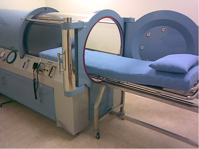
Insofar as many (perhaps ultimately all) of the cases of death are ultimately due to various forms of oxygen deficit, a powerful means of life extension may be by improving oxygen supply. One form of oxygen supply can be in the form of Hyperbaric Oxygenation (using Oxygen Pressure Chamber or Barochamber). Such therapy was indicated as beneficial against a variety of life-threatening acute conditions, such as traumas and injuries, including severe brain and chest injuries, and could also improve a variety of degenerative and aging-related conditions, from neurological impairments, such as cerebral palsy and strokes, to diabetes.[9]

However, in high concentrations, oxygen can be cytotoxic. On the positive side, oxygen toxicity against bacteria may partly explain beneficial effects of hyperbaric oxygenation for treating acute infections, as well as wound, burn and fracture healing, where infections can be a major obstacle to effective healing.[10] The cytotoxic effects may be also involved in anti-cancer treatment, to destroy cancer cells by high oxygen concentrations.[11] Yet, the oxygen cytotoxic effect can be problematic, when either the oxygen dose or time of exposure to oxygen exceed a desirable physiological threshold.[12] Oxygen can lead to excessive cell and tissue stimulation, exhausting cell replicative potential, increasing the amount of reactive oxygen species and hastening the transition to apoptotic cell death via mitochondria activation.[13] The thresholds of oxygen toxicity have been uncertain even regarding single time applications of hyperbaric oxygenation.[14] And long-term effects of hyperbaric oxygenation on human life span apparently have not been studied.


Mechanisms of action of hyperbaric oxygenation against aging-related conditions

The mechanisms of potential general protective and/or anti-aging (geroprotective) effects of hyperbaric oxygenation, if such are indeed present, are yet to be elucidated. The question of dosages may be also crucial for determining the mechanisms of such anti-aging effects. High dosages of hyperbaric oxygen should intuitively induce oxidative stress, with high reactive oxygen species (ROS) production, which has been long seen as one the major sources of molecular damage in aging.[6] Indeed, oxidative damage has been observed under hyperbaric oxygen treatment, among other effects potentially contributing to cataract development.[15] Yet, it is also appreciated that, at certain levels, ROS may stimulate tissue regeneration.[16] And yet at certain dosages, hyperbaric oxygen may produce stimulatory “hormetic” effects (i.e. stimulation by a low dose, as opposed to inhibition by a high dose of the same factor), which may in fact increase anti-oxidant protection, via stimulation of anti-oxidant defense systems.[17] The protective effects of hyperbaric oxygen by stimulating heat shock protein expression[18] and stem cell mobilization[19] have also been suggested. In a related way, chronic systemic inflammation has been long implicated as a major source of aging-related damage.[20] Furthermore, excessive neuro-inflammation has been a sustained therapeutic target.[21] Hyperbaric oxygenation has been commonly reported to produce an anti-inflammatory effect, which has been suggested as one of its major therapeutic mechanisms, for both age-related chronic and acute conditions (like acute ischemic stroke).[22] Yet, there is also a growing realization that pro-inflammatory effects may be essential for tissue regeneration, including neuro-regeneration.[23]

Closely related to the phenomenon of hormesis (low dose stimulation), hyperbaric oxygenation may exert protective anti-ischemic effects through ischemic preconditioning, that is applying a certain sub-threshold dosage of hyperbaric oxygen that would induce a transient, mild ischemia that would confer tolerance to subsequent, more severe ischemia.[24] This mechanism opens the possibility for using hyperbaric oxygenation as a preventive therapy for the elderly. Some of the mechanisms of preconditioning were associated with enhanced expression of protective enzymes, such as Sirtuins,[25] enhanced Mitogen-activated protein kinases (MAPKs) and autophagy[26] and inhibiting the mTOR pathway.[27] Thus, the mechanisms of preconditioning by hyperbaric oxygenation may be similar with the application of other geroprotective medicines (e.g. Sirtuin-stimulating or mTOR inhibiting drugs),[28] producing a general improvement of energy metabolism, yet potentially with fewer pharmacogenic side effects. However, this possibility will yet require extensive investigation, necessitating a very careful consideration of the dosages. The short vs. long term effects should be considered when studying the mechanisms of action of hyperbaric oxygenation.[29] Indeed, there may be a need for a systemic, long term evaluation of oxygen therapy effects, as a part of a whole-organism whole-life-course model of energy resources expenditures. Such models are currently only emerging,[30] and are in great need of elaboration, both experimental and theoretical.


Supplementing Hyperbaric Oxygenation with additional treatment modalities

Hyperbaric Oxygenation can be seen as a potentially effective therapeutic or stimulating means, but it is unclear to which extent it can serve as a lifespan-extending means. Hence, the therapeutic modality of Hyperbaric Oxygenation may be supplemented or followed by additional modalities particularly designed to serve life-prolonging, rejuvenative and reparative functions. Pressure chamber could in principle provide a convenient environment to implement such modalities, insofar as it provides a protected, isolated and enclosed space unit, which can be easily manipulated and monitored for a variety of purposes. Some of the potential reparative applications can be as follows.


O2/CO2 Balance

For the life-span and health-span extension, rather than applying hyperbaric oxygenation, actually reducing oxygen partial pressure may be beneficial. Thus people living at high altitudes (with reduced oxygen pressure) are noted for high longevity, presumably due to either reduced metabolic rate or long-term adaptations. Some of the potential beneficial adaptations may include increased production of red blood cells, formation of new capillaries and increase in respiratory enzymes, and other mechanisms.[31]

Also, increased CO2 can be beneficial, insofar as persistent hypercapnia (enhanced CO2 level) has been associated with an increased life-span in animal models. This may presumably be due to increasing blood alkalinity (through liberating bicarbonate reserves) which may in turn positively affect proteins’ isoelectric stability.[32]

Interestingly and seemingly paradoxically, both hyperbaric oxygenation therapy and its apparent opposite – therapeutic hypoxia or hypoxic training (for example intermittent hypoxia) have been suggested to produce positive preconditioning effects against ischemic aging-related conditions, such as heart disease and neurodegenerative diseases.[33] The apparent paradox may be once again explained by the phenomenon of “hormesis” – namely the activation of anti-hypoxic/anti-ischemic protective mechanisms by certain extents of both deficit and excess of oxygen (in the latter case possibly increasing reactive oxygen species levels to induce a protective counter-effect), as well as possibly by other mild stressors (chemical, mechanical or electrical). The precise dosages and thresholds of such similar protective effects by seemingly diverse means, as well as their potential common central neuro-humoral regulatory mechanisms, yet require elucidation.

Besides hyperbaric oxygen therapy, normobaric (normal pressure) oxygen therapy, or just oxygen therapy generally (increasing oxygen supply) has been a widely applied means of therapy and resuscitation.[34] While hyperbaric oxygenation (using a pressure chamber) may be more effective to achieve rapid oxygen delivery to deep vital tissues, normobaric oxygenation (e.g. using an oxygen mask) may be more conveniently applicable and less expensive. Yet for normobaric oxygenation too, the appropriate balance of O2/CO2 levels may be critical. The pressure chamber may provide an ideal environment to control both O2 and CO2 levels and pressures, for acute therapeutic or prolonged restorative regimens. Still, with regard to CO2 manipulation, its long term effects as well as its effects on immediate daily performance will yet need to be established.

The issue of optimal thresholds or O2/CO2 balance will be vital, insofar as excessive O2 application may lead to a “burnout,” while excessive CO2 application may lead to a “death zone.” Both acidosis and alkalosis may be produced by O2/CO2 imbalance. Perhaps the most beneficial therapeutic regimen may be maintaining and/or rapidly restoring the physiological O2/CO2 balance. The normal (balanced) concentration of alveolar CO2 is often assumed to be about 6.5%, yet may vary according to particular metabolic requirements of every individual.[35]


Monitoring

In order to personalize the therapy, and to ensure its safety and efficacy, the treatment modalities should be related with a thorough array of monitoring and evaluation modalities, in particular for the evaluation of the organism’s energy metabolism, before, during and after the treatment. In performing oxygen therapy (in particular hyperbaric oxygenation), reference need to be made for Oxygen and CO2 balance (supply vs. demand), as well as for the supply and demand of macroergic (energy-rich) substances, in the entire organism and particular organs.[36] Measurement modalities may include oxygen measurement by mitochondrial cytochrome a,a3 reflectance spectrophotometry, mitochondrial NADH redox state by NADH fluorometry, tissue blood flow by Laser Doppler Flowmetry, hemoglobin oxygen saturation by reflectometry, DC potential and various ionic levels by micro-electrodes, gas partial pressure (O2, CO2, NO, etc) and pH levels by micro-electrodes and optodes, up to more advanced methods such as functional magnetic resonance imaging (fMRI) or using sequential single photon emission computerized tomography (SPECT) scans, etc.[37]

The monitoring of the gas composition (PaO2 and PaCO2) of the arterial blood may be seen as a necessary condition to perform effective hyperbaric oxygen therapy against life-threatening situations in patients with deteriorating cardiorespiratory functions, especially for the elderly patients.[8] This is necessary in order to control and maintain adequate levels of lung gas exchange, with regulated parameters of oxygen supply and controlled oxygen concentration. The speed of the blood flow and blood pressure in vital organs are also among the critical vital signs that need to be known. The basic parameters of cardiorespiratory function need to be monitored, such as: PaO2 mmHg – arterial partial pressure of oxygen; PaCO2 – arterial partial pressure of CO2; PaO2/FiO2 – the ratio of the partial pressure of oxygen to the fraction of inspired oxygen; SVI ml/m2 – stroke volume index; CI L/min/m2 – cardiac index; pH – blood acidity level. Such measures of cardio-respiratory function can help reference the normal balanced O2/CO2 levels in the blood and favorable blood electric charge and hemodynamic conditions.

The anatomical and physiological effects of aging on the heart and lungs are also vitally important parameters, including such indicators as the rise of arterial pressure and resistance with aging due to the increasing arterial stiffness, reduced contractility and relaxation of the heart, a reduction of lung vital capacity, impairment of gas mixture in the lungs, and other harmful anatomical and physiological effects of aging. All the deteriorative changes in the cardio-respiratory system generally show in the reduction of maximal oxygen uptake (VO2max) which has been considered one of the most informative parameters for biological age evaluation.[38]

The above parameters of cardiorespiratory function are practically indispensable in emergency and intensive care medicine, when treating acute and often life-threatening conditions. Yet, arguably, the cardio-respiratory parameters routinely employed in emergency and intensive care medicine may be good candidates for biomarkers of aging as they have proved their utility as real-time indicators of the organism’s vitality and energy. Often, in general frailty assessments, energy levels in the elderly are evaluated simply by asking the question “Do you feel full of energy?”[39] Yet, there may be more objective measures of the aging organism’s energy level, by such means as spirometry, oximetry, hemodynamic, electrochemical and spectroscopic energy metabolite measurements, as well as other structural and functional parameters of the cardiorespiratory system, that can provide improved indication for therapy.[40] Arguably, such cardio-respiratory “physiomic” parameters or markers of aging may be clinically valuable and conveniently interpretable for a practicing physician, alongside the many “biomarkers of aging” based on predominantly molecular-biological, e.g. genetic, epigenetic and other “omic” age-related alterations that are currently investigated. [41]

The main clinical utility of biomarkers or diagnostic parameters of aging is that their changes can help evaluate the effectiveness of particular therapeutic regimens, especially the effectiveness of particular therapeutic dosages. Yet curiously, in hyperbaric oxygen therapy, the concept of dosage is only rudimentary and there is no commonly agreed way to define the dosage. Moreover, there is no agreed way to evaluate the effects of this kind of therapy, and correspondingly no agreed way to correlate between the dose and the effect. The same may be said regarding other potential “energy-modulating” interventions into aging, whose definition is yet very nebulous. In certain studies, it was suggested to define the dose of hyperbaric oxygen therapy as the product of intra-barochamber pO2 (ATA), the duration of a single hyperbaric oxygenation exposure (hours), and the number of hyperbaric oxygenation treatments, yielding the dose unit: (ATA*h*N).[8,12] In those studies, the efficacy of hyperbaric oxygenation therapy was evaluated according to the number of patients who showed a significant clinical improvement in their neurological state in the course of the treatment (the percent of the total number of patients). The level of the therapy efficacy was compared with a corresponding value of the dose. For the treatment of acute ischemic stroke, a higher efficacy was indicated with increasing the average total hyperbaric oxygenation dose, reaching the maximum efficacy with the average doses of no less than 30 units (ATA*h*N).[12] However, such a definition of the dosages has not become consensus, and the definition of the therapy effects and of the dose-effect relations are rather vague and yet require a thorough elaboration and clarification. Hopefully, thanks to refinement of the definitions and massive additional data collection on dose-effect relations, including the evaluation of long-term effects and differential personalized effects in different patient groups (e.g. the elderly vs. the young) – oxygen therapy, hyperbaric oxygen therapy particularly, or “energy-modulating therapy” more generally – can become efficient means to alleviate aging-related conditions and increase healthy and productive life.

It is also necessary to note, that the obtained datasets of biomarkers, diagnostic parameters, and dose-effect relations, will be not only necessary for monitoring and personalizing treatment regimens, but will also be able to provide invaluable information for many yet unforeseen “quantified health” and “quantified longevity” applications – collecting a vast amount of health data on aging-related changes and their possible improvements to enable planning better informed therapeutic and life-style regimens and strategies to achieve healthy longevity.


Comprehensive physiological manipulation unit

Enhancing blood supply: The main purpose of oxygenation therapy (in particular hyperbaric oxygen therapy) is to directly enhance the supply of potentially deficient oxygen to the tissues that need it (while necessarily watching out against “burning out” and “oxygen toxicity”). However, oxygen supply can be improved by more indirect means, such as improving blood supply to the tissues. Historically, improved blood supply to the tissues (also for the purposes of rejuvenation) has been persistently sought. For example, the whole-body increase of the blood flow (hyperemia) has been achieved by various means ranging from hormone replacement therapy (by supplements, tissue transplants, and even operations on the endocrine organs) through diathermy (tissue heating), massage, exercise and baths.[1] The problems of oxygen delivery to the vital tissues have been also tackled from additional angles, such as oxygenated microparticles and “artificial blood”,[42] various forms of heart-lung machines, artificial hearts and other assisted circulation devices,[43] or pharmacological means to improve energy metabolism.[28] The blood flow can be also stimulated by electromagnetic devices.

As briefly mentioned above, recording electrodes and magnetic resonance devices may be employed for monitoring (for example during the course of oxygen therapy). Yet, in addition, stimulation electrodes may be also used for physiological manipulation purposes, in particular to stimulate nervous activity and blood flow, even to stimulate tissue regeneration, in particular blood vessels growth (angiogenesis – another potential means to improve tissue blood supply and oxygenation, but also requiring caution to avoid uncontrolled growth).[44] Such electromagnetic therapeutic devices have been sometimes termed “electroceuticals.”[45] They can be incorporated into the therapeutic regimens, either within the pressure chamber or as a part of accompanying regimens.

The incorporation or fitting of the additional therapeutic modalities within the pressure chamber can provide additional benefits. The ability provided by the pressure chamber to control and manipulate pressure, gas concentrations and temperature, can produce a convenient environment for physiological manipulation. Furthermore, with the addition of an infusion apparatus for delivering medications, including various regenerative and anti-aging medications (depending on the costs involved), this can become a multifunctional treatment unit. Some of its functions can be as follows.

“Resting state” induction: One possibility may be inducing a restorative “resting state” through a variety of physical means (pressure, temperature, oxygen and carbon dioxide concentration) as well as by electromagnetic and pharmacological means. Thus both reversible hypothermic and pharmacological resting states (reversible coma) are already becoming widely used clinical methods for recuperation and resuscitation, and can be incorporated into the chamber.[46] Just by using such physical manipulation means that are available in the pressure chamber – such as pressure, temperature, O2 and CO2 concentrations – a resting state can be induced insofar as rest and sleep are characterized by particular breathing and temperature patterns.[47] Temperature control can be another powerful means of physical manipulation. Thus, lower core body temperature has been correlated with longer lifespans.[48] In particular, lowering the body temperature (hypothermia) during hyperbaric oxygenation treatment could reduce energy (oxygen) demand by the organism and thus potentially lower oxygen toxicity. On the other hand, increased blood flow through heating may be used for therapeutic stimulation purposes. Within the chamber, temperature can be manipulated in both directions. The electric charge of the breathing mixture (e.g. negative ionization) can be also significant for recuperation vs. stimulation.[49]

Sleep enhancement: Sleep enhancement can be yet another promising restorative modality (in fact a form of “resting state”). In particular, slow-wave sleep (Stage 3, with synchronized EEG activity, showing slow waves with a frequency of less than 1 Hz) has been known to be vital for recuperation, presumably due to enhanced growth hormone production[50] or synchronization of physiological functions. [51] The restorative effects of sleep generally, and deep sleep in particular, may be also possibly due to activation of the immune response during sleep, or elimination of toxins, or other mechanisms.[52] This stage can be induced by a variety of methods, including: transcranial direct current stimulation (tDCS) and transcranial magnetic stimulation (TMS),[53] other forms of sensory sleep stimulation[54] and a variety of slow-wave sleep enhancing drugs.[55] Sleeping in a hyperbaric chamber has already been practiced, and this combination of therapeutic modalities can be further explored and expanded if proven safe and effective.

All such technologies are yet extremely experimental, dose responses have not been thoroughly studied, hence side effects may be unpredictable. Both their short and long term effects are largely unknown, and the introduction of such additional technologies may become prohibitively expensive and unwieldy. They are mentioned here only as possibilities that can be further investigated and prospectively included within a potential “comprehensive restorative chamber” or “survival chamber.”


References and notes

[1] Ilia Stambler, A History of Life-Extensionism in the Twentieth Century, Longevity History, 2014, http://www.longevityhistory.com/.

[2] Ilia Stambler, “Human life extension: opportunities, challenges, and implications for public health policy,” in: Alexander Vaiserman (Ed.), Anti-aging Drugs: From Basic Research to Clinical Practice, Royal Society of Chemistry, London, 2017, pp. 535-564, http://pubs.rsc.org/en/content/ebook/978-1-78262-435-6#!divbookcontent;

Alexander Vaiserman, Oleh Lushchak, “Anti-aging drugs: where are we and where are we going?” in: Alexander Vaiserman (Ed.), Anti-aging Drugs: From Basic Research to Clinical Practice, Royal Society of Chemistry, London, 2017, pp. 3-10, http://pubs.rsc.org/en/content/ebook/978-1-78262-435-6#!divbookcontent.

[3]. Anthony Atala, “Extending life using tissue and organ replacement,” Current Aging Science, 1(2), 73-83, 2008, http://www.eurekaselect.com/95101/article;

Giuseppe Orlando, Shay Soker, Robert J. Stratta, Anthony Atala, “Will Regenerative Medicine Replace Transplantation?” Cold Spring Harbor Perspectives in Medicine, 3(8), a015693, 2013, https://www.ncbi.nlm.nih.gov/pmc/articles/PMC3721273/.

[4] Ronald Bellamy, Peter Safar, Samuel Tisherman, …, Harvey Zar, “Suspended animation for delayed resuscitation,” Critical Care Medicine, 24(2Suppl), S24-47, 1996, http://www.ncbi.nlm.nih.gov/pubmed/8608704.

[5] Yury P. Gerasimenko, Daniel C. Lu, Morteza Modaber, …, V. Reggie Edgerton, “Noninvasive Reactivation of Motor Descending Control after Paralysis,” Journal of Neurotrauma, 32(24), 1968-1980, 2015, http://online.liebertpub.com/doi/abs/10.1089/neu.2015.4008;

Max Schaldach, Electrotherapy of the Heart: Technical Aspects in Cardiac Pacing, Springer-Verlag, Berlin, 2012.

[6] Denham Harman, “Aging: a theory based on free radical and radiation chemistry,” Journal of Gerontology, 11, 298-300, 1956, http://www.uccs.edu/Documents/rmelamed/harman_1956_13332224.pdf;

Rajindar S. Sohal, “Role of oxidative stress and protein oxidation in the aging process,” Free Radical Biology and Medicine, 33(1), 37-44, 2002, http://www.sciencedirect.com/science/article/pii/S0891584902008560;

Rajindar S. Sohal, “Oxidative stress hypothesis of aging,” Free Radical Biology and Medicine, 33(5), 573-574, 2002, http://www.sciencedirect.com/science/article/pii/S0891584902008857;

Toren Finkel, Nikki J. Holbrook, “Oxidants, oxidative stress and the biology of ageing,” Nature, 408(6809), 239-247, 2000, https://www.nature.com/nature/journal/v408/n6809/full/408239a0.html.

[7] Gerald J. Gruman, A History of Ideas about the Prolongation of Life. The Evolution of Prolongevity Hypotheses to 1800, Transactions of the American Philosophical Society, Vol. 56 (9), Philadelphia, 1966.

[8] Gennady G. Rogatsky, Ilia Stambler, “Hyperbaric oxygenation for resuscitation and therapy of elderly patients with cerebral and cardio-respiratory dysfunction,” Frontiers In Bioscience (Scholar Edition), 9, 230-243, June 1, 2017, http://www.bioscience.org/2017/v9s/af/484/2.htm, https://www.bioscience.org/special-issue-details?editor_id=1746, https://www.ncbi.nlm.nih.gov/pubmed/28410116.

[9] The standard indications for the use of hyperbaric oxygenation, as established by the US-incorporated Undersea and Hyperbaric Medicine Society (UHMS), include: 1. Air or Gas Embolism, 2. Carbon Monoxide Poisoning, 3. Clostridial Myositis and Myonecrosis (Gas Gangrene), 4. Crush Injury, Compartment Syndrome and Other Acute Traumatic Ischemias, 5. Decompression Sickness, 6. Arterial Insufficiencies, 7. Severe Anemia, 8. Intracranial Abscess, 9. Necrotizing Soft Tissue Infections, 10. Osteomyelitis (Refractory), 11. Delayed Radiation Injury (Soft Tissue and Bony Necrosis), 12. Compromised Grafts and Flaps, 13. Acute Thermal Burn Injury; 14. Idiopathic Sudden Sensorineural Hearing Loss.

(Undersea and Hyperbaric Medicine Society (UHMS), “Indications for Hyperbaric Oxygen Therapy,” https://www.uhms.org/resources/hbo-indications.html.)

Yes, there is good evidence for the possible use of this treatment against other severe and chronic conditions. See for example:

Gennady G. Rogatsky, Avraham Mayevsky, “The life-saving effect of hyperbaric oxygenation during early-phase severe blunt chest injuries,” Undersea and Hyperbaric Medicine, 34(2), 75-81, 2007, http://archive.rubicon-foundation.org/xmlui/bitstream/handle/123456789/6468/17520858.pdf?sequence=1;

Ning Gu, Fumiko Nagatomo, Hidemi Fujino, Isao Takeda, Kinsuke Tsuda, Akihiko Ishihara, “Hyperbaric oxygen exposure improves blood glucose level and muscle oxidative capacity in rats with type 2 diabetes,” Diabetes Technology & Therapeutics, 12(2), 125-133, 2010, http://online.liebertpub.com/doi/abs/10.1089/dia.2009.0104;

Majid Kalani, Gun Jörneskog, Nazanin Naderi, Folke Lind, Kerstin Brismar, “Hyperbaric oxygen (HBO) therapy in treatment of diabetic foot ulcers: Long-term follow-up,” Journal of Diabetes and its Complications, 16(2), 153-158, 2002, http://www.jdcjournal.com/article/S1056-8727(01)00182-9/fulltext .

Michael H. Bennett, Jan P. Lehm, Nigel Jepson, “Hyperbaric oxygen therapy for acute coronary syndrome,” Cochrane Database of Systematic Reviews, 2015(7), CD004818, 2015, http://onlinelibrary.wiley.com/doi/10.1002/14651858.CD004818.pub4/full;

Peter Kranke, Michael H. Bennett, Marrissa Martyn-St James, Alexander Schnabel, Sebastian E. Debus, “Hyperbaric oxygen therapy for chronic wounds,” Cochrane Database of Systematic Reviews, 2015(6), CD004123, 2015, http://onlinelibrary.wiley.com/doi/10.1002/14651858.CD004123.pub3/abstract;

Michael H Bennett, Barbara Trytko, Benjamin Jonker, “Hyperbaric oxygen therapy for the adjunctive treatment of traumatic brain injury,” Cochrane Database of Systematic Reviews, 2012(12), CD004609, 2012, http://onlinelibrary.wiley.com/doi/10.1002/14651858.CD004609.pub2/abstract;

Richard A. Neubauer Research Institute, “Resources,” www.ranri.org/resources.html.

[10] Escobar S.J., Slade J.B., Hunt T.K., Cianci P., “Adjuvant hyperbaric oxygen therapy (HBO2) for treatment of necrotizing fasciitis reduces mortality and amputation rate,” Undersea and Hyperbaric Medicine, 32(6), 437–43, 2005, http://dspace.rubicon-foundation.org/xmlui/bitstream/handle/123456789/4061/16509286.pdf?sequence=1.

[11] Ingrid Moen, Linda E. B. Stuhr, “Hyperbaric oxygen therapy and cancer – a review,” Targeted Oncology, 7, 233–242, 2012, https://www.ncbi.nlm.nih.gov/pmc/articles/PMC3510426/.

[12] Gennady G. Rogatsky, Edward G. Shifrin, Avraham Mayevsky, “Optimal dosing as a necessary condition for the efficacy of hyperbaric oxygen therapy in acute ischemic stroke: a critical review,” Neurological Research, 25(1), 95-98, 2003, http://www.tandfonline.com/doi/abs/10.1179/016164103101201003; Optimal_dosing_as_a_necessary_condition_for_the_efficacy_

of_hyperbaric_oxygen_therapy_in_acute_ischemic_stroke_A_critical_review.

[13] Elena Afrimzon, Naomi Zurgil, Yana Shafran, Pnina Leibovich, Maria Sobolev, Larissa Guejes, Mordechai Deutsch, “The use of sequential staining for detection of heterogeneous intracellular response of individual Jurkat cells to lysophosphatidylcholine,” Journal of Immunological Methods, 387(1-2), 96-106, 2013, http://www.sciencedirect.com/science/article/pii/S0022175912002992.

[14] Efouni S.N., et al., “Hyperoxia. Patofiziologicheskie aspekty lechebnogo i toxicheskogo vozdeystvia hyperbaricheskogo kisloroda (Hyperoxia. Pathophysiological aspects of therapeutic and toxic effects of hyperbaric oxygenation), in: EShfouni S.N. (Ed.), Rukovodstvo po Hyperbaricheskoy Oxygenazii. Teoria I Praktika Klinicheskogo Primenenia (Hyperbaric Oxygenation: a manual. The theory and practice of clinical application), Akademia Medizinskikh Nauk SSSR (Academy of Medical Sciences of the USSR), Medizina, Moscow, 1986, pp. 29-56;

Richard D. Vann, Oxygen Toxicity Risk Assessment,   Office of Naval Research, Arlington VA, 1988, https://www.researchgate.net/publication/235051634_Oxygen_Toxicity_Risk_Assessment;

Noemi Bitterman, “CNS oxygen toxicity,” Undersea and Hyperbaric Medicine, 31 (1), 63-72, 2004, http://dspace.rubicon-foundation.org/xmlui/bitstream/handle/123456789/3991/15233161.pdf?sequence=1.

[15] Yi Zhanga, Shan OuYanga, Lan Zhangb, XianLing Tanga, Zhen Songa, Ping Liua, “Oxygen-induced changes in mitochondrial DNA and DNA repair enzymes in aging rat lens,” Mechanisms of Ageing and Development, 131(11-12), 666-673, 2010, http://www.sciencedirect.com/science/article/pii/S0047637410001740.

[16] Carole Gauron, Christine Rampon, Mohamed Bouzaffour, Eliane Ipendey, Jérémie Teillon, Michel Volovitch, Sophie Vriz, “Sustained production of ROS triggers compensatory proliferation and is required for regeneration to proceed,” Scientific Reports, 3, 2084, 2013, https://www.nature.com/articles/srep02084.

[17] Cassandra A. Godman, Rashmi Joshi, Charles Giardina, George Perdrizet, Lawrence E. Hightower, “Hyperbaric oxygen treatment induces antioxidant gene expression,” Annals of the New York Academy of Sciences, 1197, 178-183, 2010, http://onlinelibrary.wiley.com/doi/10.1111/j.1749-6632.2009.05393.x/abstract.

[18] Jeysen Zivan Yogaratnam, Gerard Laden, Levant Guvendik, Mike Cowen, Alex Cale, Steve Griffin, “Can hyperbaric oxygen be used as adjunctive heart failure therapy through the induction of endogenous heat shock proteins?” Advances in Therapy, 24(1), 106-118, 2007, https://oxfordrecoverycenter.com/wp-content/uploads/2017/02/Can-Hyperbaric-Oxygen-Be-Used-as-Adjunctive-Heart-Failure-Therapy-Through-the-Induction-of-Endogenous-Heat-Shock-Proteins.pdf.

[19] Stephen R. Thom, Veena M. Bhopale, Omaida C. Velazquez, Lee J. Goldstein, Lynne H. Thom, Donald G. Buerk, “Stem cell mobilization by hyperbaric oxygen,” American Journal of Physiology – Heart and Circulatory Physiology, 290(4), H1378-1386, 2006, http://ajpheart.physiology.org/content/290/4/H1378.long.

[20] Claudio Franceschi, Judith Campisi, “Chronic inflammation (inflammaging) and its potential contribution to age-associated diseases,” The Journals of Gerontology Series A, Biological Sciences and Medical Sciences, 69(Suppl. 1), S4-9, 2014, https://academic.oup.com/biomedgerontology/article-lookup/doi/10.1093/gerona/glu057.

[21] Joaquin Jordan, Tomas Segura, David Brea, Maria F. Galindo, Jose Castillo, “Inflammation as therapeutic objective in stroke,” Current Pharmaceutical Design, 14, 3549-3564, 2008, http://www.eurekaselect.com/68189/article.

[22] Zheng Ding, Wesley C. Tong, Xiao-Xin Lu, Hui-Ping Peng, “Hyperbaric oxygen therapy in acute ischemic stroke: a review,” Interventional Neurology, 2(4), 201-211, 2014, https://www.ncbi.nlm.nih.gov/pmc/articles/PMC4188156/.

[23] Michael Karin, Hans Clevers, “Reparative inflammation takes charge of tissue regeneration,” Nature, 529(7586), 307-315, 2016, http://www.nature.com/nature/journal/v529/n7586/full/nature17039.html.

[24] Qin Hu, Anatol Manaenko, Nathanael Matei, Zhenni Guo, Ting Xu, Jiping Tang, John H. Zhang, “Hyperbaric oxygen preconditioning: a reliable option for neuroprotection,” Medical Gas Research, 6(1), 20-32, 2016, https://www.ncbi.nlm.nih.gov/pmc/articles/PMC5075679/;

Zheng Ding, Wesley C. Tong, Xiao-Xin Lu, Hui-Ping Peng, “Hyperbaric oxygen therapy in acute ischemic stroke: a review,” Interventional Neurology, 2(4), 201-211, 2014, https://www.ncbi.nlm.nih.gov/pmc/articles/PMC4188156/;

Gerd Heusch, Hans Erik Bøtker, Karin Przyklenk, Andrew Redington, Derek Yellon, “Remote Ischemic Conditioning,” Journal of the American College of Cardiology, 65(2), 177-195, 2015, https://www.ncbi.nlm.nih.gov/pmc/articles/PMC4297315/.

[25] Wenjun Yan, Zongping Fang, Qianzi Yang, Hailong Dong, Yan Lu, Chong Lei, Lize Xiong, “SirT1 mediates hyperbaric oxygen preconditioning-induced ischemic tolerance in rat brain,” The Journal of Cerebral Blood Flow & Metabolism, 33(3), 396-406, 2013, https://www.ncbi.nlm.nih.gov/pmc/articles/PMC3587810/.

[26] Xiao-qian Liu, Rui Sheng, Zheng-hong Qin, “The neuroprotective mechanism of brain ischemic preconditioning,” Acta Pharmacologica Sinica, 30(8), 1071-1080, 2009, https://www.ncbi.nlm.nih.gov/pmc/articles/PMC4006675/.

[27] Mikhail V. Blagosklonny, “Hormesis does not make sense except in the light of TOR-driven aging,” Aging (Albany NY), 3(11), 1051-1062, 2011, http://www.aging-us.com/article/100411/text.

[28] Valter D. Longo, Adam Antebi, Andrzej Bartke, Nir Barzilai, Holly M. Brown-Borg, Calogero Caruso, Tyler J. Curiel, Rafael de Cabo, Claudio Franceschi, David Gems, Donald K. Ingram, Thomas E. Johnson, Brian K. Kennedy, Cynthia Kenyon, Samuel Klein, John J. Kopchick, Guenter Lepperdinger, Frank Madeo, Mario G. Mirisola, James R. Mitchell, Giuseppe Passarino, Karl L. Rudolph, John M. Sedivy, Gerald S. Shadel, David A. Sinclair, Stephen R. Spindler, Yousin Suh, Jan Vijg, Manlio Vinciguerra, Luigi Fontana, “Interventions to slow aging in humans: Are we ready?” Aging Cell, 14(4), 497-510, 2015, http://www.ucl.ac.uk/~ucbtdag/Longo_2015.pdf;

Ilia Stambler, “Stop Aging Disease! ICAD 2014,” Aging and Disease, 6(2), 76-94, 2015, http://www.aginganddisease.org/EN/10.14336/AD.2015.0115;

Ilia Stambler, “Human life extension: opportunities, challenges, and implications for public health policy,” in: Alexander Vaiserman (Ed.), Anti-aging Drugs: From Basic Research to Clinical Practice, Royal Society of Chemistry, London, 2017, pp. 535-564, http://pubs.rsc.org/en/content/ebook/978-1-78262-435-6#!divbookcontent;

Alexander Vaiserman, Oleh Lushchak, “Anti-aging drugs: where are we and where are we going?” in: Alexander Vaiserman (Ed.), Anti-aging Drugs: From Basic Research to Clinical Practice, Royal Society of Chemistry, London, 2017, pp. 3-10, http://pubs.rsc.org/en/content/ebook/978-1-78262-435-6#!divbookcontent.

[29] Liran I. Shlush, Karl L. Skorecki, Shalev Itzkovitz, Shiran Yehezkel, Yardena Segev, Hofit Shachar, Ron Berkovitz, Yochai Adir, Irma Vulto, Peter M. Lansdorp, Sara Selig, “Telomere elongation followed by telomere length reduction, in leukocytes from divers exposed to intense oxidative stress – implications for tissue and organismal aging,” Mechanisms of Ageing and Development, 132(3), 123-130, 2011, http://www.sciencedirect.com/science/article/pii/S0047637411000224.

[30] Vasilij N. Novoseltsev, Janna Novoseltseva, Anatoli I. Yashin, “A homeostatic model of oxidative damage explains paradoxes observed in earlier aging experiments: a fusion and extension of older theories of aging,” Biogerontology, 2(2), 127-138, 2001, https://link.springer.com/article/10.1023/A:1011511100472.

[31] John B. West, “Exciting Times in the Study of Permanent Residents of High Altitude,” High Altitude Medicine & Biology, 12 (1), 1, 2011, http://online.liebertpub.com/doi/abs/10.1089/ham.2011.12101;

Martin Burtscher, “Effects of Living at Higher Altitudes on Mortality: A Narrative Review,” Aging and Disease, 5(4), 274–280, 2014, https://www.ncbi.nlm.nih.gov/pmc/articles/PMC4113517/;

Jay F. Storz, Hideaki Moriyama, “Mechanisms of Hemoglobin Adaptation to High Altitude Hypoxia,” High Altitude Medicine & Biology, 9(2), 148-157, 2008, https://www.ncbi.nlm.nih.gov/pmc/articles/PMC3140315/;

Jean-Marie Robine, Fred Paccaud, “Nonagenarians and centenarians in Switzerland, 1860–2001: a demographic analysis,” Journal of Epidemiology and Community Health, 59(1), 31-37, 2005, http://jech.bmj.com/content/59/1/31;

Alexander Leaf, Youth in Old Age, McGraw-Hill Book Company, New York, 1975;

Khatchik Muradian, “Atmosphere, Metabolism and Longevity,” in: Alexander M. Vaiserman, Alexey A. Moskalev, Elena G. Pasyukova (Eds.), Life Extension: Lessons from Drosophila, Dordrecht, Springer, 2015.

[32] Khachik Muradian, “’Pull and push back’ concepts of longevity and life span extension,” Biogerontology, 14(6), 687-691, 2013, https://link.springer.com/article/10.1007%2Fs10522-013-9472-1;

Reuven Tirosh, “Ballistic Protons and Microwave-induced Water Solitons in Bioenergetic Transformations,” International Journal of Molecular Sciences, 7(9), 320-345, 2006, http://www.mdpi.com/1422-0067/7/9/320.

[33] Angela Navarrete-Opazo, Gordon S. Mitchell, “Therapeutic potential of intermittent hypoxia: a matter of dose,” American Journal of Physiology – Regulatory, Integrative and Comparative Physiology, 307(10), R1181–1197, 2014, http://ajpregu.physiology.org/content/307/10/R1181.

[34] Michael H. Bennett, Christopher French, Alexander Schnabel, Jason Wasiak, Peter Kranke, Stephanie Weibel, “Normal pressure oxygen therapy and hyperbaric oxygen therapy for migraine and cluster headaches,” The Cochrane Database of Systematic Reviews, (12):CD005219, 2015, http://onlinelibrary.wiley.com/doi/10.1002/14651858.CD005219.pub3/full;

Aneesh B. Singhal, “A review of oxygen therapy in ischemic stroke,” Neurological Research, 29(2), 173-183, 2007, http://www.tandfonline.com/doi/abs/10.1179/016164107X181815.

[35] Konstantin Pavlovich Buteyko, Metod Buteyko. Opyt Vnedrenia v Medizinskuyu Praktiku (The Buteyko Method. The Experience of Application in Medical Practice), Patriot, Moscow, 1990, http://www.buteyko.ru/rus/index.shtml;

Vladimir K. Buteyko and Marina M. Buteyko, The Buteyko Theory about a Key Role of Breathing for Human Health, Scientific Introduction to the Buteyko Therapy for Experts (in Russian and English), Buteyko Co LTD, Voronezh, 2005, http://www.doctorbuteykodiscoverytrilogy.com/the-buteyko-theory-about-a-key-role-of-breathing-for-human-health.php;

Paul M Macey, Mary A Woo, Ronald M Harper, “Hyperoxic Brain Effects Are Normalized by Addition of CO2,” PLoS Medicine, 4(5), e173, 2007, https://doi.org/10.1371/journal.pmed.0040173.

[36] Gennady G. Rogatsky, Edward G. Shifrin, Avraham Mayevsky, “Physiologic and biochemical monitoring during hyperbaric oxygenation,” Undersea & Hyperbaric Medicine, 26(2), 111-122, 1999, Physiologic_and_biochemical_monitoring_during_hyperbaric_oxygenation.

[37] Judith Sonn, Avraham Mayevsky, “Responses to Cortical Spreading Depression under Oxygen Deficiency,” The Open Neurology Journal, 6, 6-17, 2012, https://www.ncbi.nlm.nih.gov/pmc/articles/PMC3367297/;

Greg Zaharchuk, Reed F. Busse, Guy Rosenthal, Geoffery T. Manley, Orit A. Glenn, William P. Dillon, “Noninvasive oxygen partial pressure measurement of human body fluids in vivo using magnetic resonance imaging,” Academic Radiology, 13(8), 1016-24, 2006, http://www.sciencedirect.com/science/article/pii/S1076633206002704;

Nili Zarchin, Sigal Meilin, Joseph Rifkind, Avraham Mayevsky, “Effect of aging on brain energy-metabolism,” Comparative Biochemistry and Physiology Part A: Molecular & Integrative Physiology, 132(1), 117-120, 2002, http://www.sciencedirect.com/science/article/pii/S1095643301005372.

[38] Ward Dean, “Biologic aging measurement: its rationale, history, and current status,” in: Arthur K. Balin (Ed.), Human Biologic Age Determination, Boca Raton FL, CRC Press, 1994, pp. 3-14;

Walter M. Bortz 4th, Walter M. Bortz 2nd, “How fast do we age? Exercise performance over time as a biomarker,” Journal of Gerontology A, Biological Sciences and Medical Sciences, 51(5), M223-225, 1996, https://www.ncbi.nlm.nih.gov/pubmed/8808993;

Johannes H.G.M. van Beek, Thomas B.L. Kirkwood, James B. Bassingthwaighte, “Understanding the physiology of the ageing individual: computational modelling of changes in metabolism and endurance,” Interface Focus, 6(2), 20150079, 2016, http://rsfs.royalsocietypublishing.org/content/6/2/20150079.

[39] Kristine E. Ensrud, Susan K. Ewing, Peggy M. Cawthon, Howard A. Fink, Brent C. Taylor, Jane A. Cauley, Thuy-Tien Dam, Lynn M. Marshall, Eric S. Orwoll, Steven R. Cummings, and The Osteoporotic Fractures in Men (MrOS) Research Group, “A comparison of frailty indexes for the prediction of falls, disability, fractures, and mortality in older men,” Journal of the American Geriatrics Society, 57(3), 492-498, 2009, https://www.ncbi.nlm.nih.gov/pmc/articles/PMC2861353/;

Linda P. Fried, Catherine M. Tangen, Jeremy Walston, Anne B. Newman, Calvin Hirsch, John Gottdiener, Teresa Seeman, Russell Tracy, Willem J. Kop, Gregory Burke, Mary Ann McBurnie, for the Cardiovascular Health Study Collaborative Research Group, “Frailty in older adults: evidence for a phenotype,” Journal of Gerontology A, Biological Sciences and Medical Sciences, 56(3), M146–M156, 2001, http://www.sld.cu/galerias/pdf/sitios/gericuba/fenotipo_frailty.pdf.

[40] Yang Guang, Dong Feng, Li Yuzhong, Liu Hui, “Theoretical and Experimental Evidence in Mixed Serum Samples as Natural Reference Materials for Measuring Activity of Biomacromolecules in Serum,” Clinical Laboratory, 64, 04/2017, https://www.clin-lab-publications.com/article/2331;

Avraham Mayevsky, Efrat Barbiro-Michaely, “Shedding light on mitochondrial function by real time monitoring of NADH fluorescence: II: human studies,” International Journal of Clinical Monitoring and Computing, 27(2), 125-145, 2013, https://link.springer.com/article/10.1007/s10877-012-9413-6.

[41] Robert N. Butler, Richard Sprott, Huber Warner, Jeffrey Bland, Richie Feuers, Michael Forster, Howard Fillit, S. Mitchell Harman, Michael Hewitt, Mark Hyman, Kathleen Johnson, Evan Kligman, Gerald McClearn, James Nelson, Arlan Richardson, William Sonntag, Richard Weindruch, Norman Wolf, “Biomarkers of aging: from primitive organisms to humans,” Journal of Gerontology A, Biological Sciences Medical Sciences, 59, B560-567, 2004, https://www.researchgate.net/publication/8493491_Aging_The_Reality_Biomarkers_of_Aging_From_Primitive_Organisms_to_Humans;

Thomas Craig, Chris Smelick, Robi Tacutu, Daniel Wuttke, Shona H. Wood, Henry Stanley, Georges Janssens, Ekaterina Savitskaya, Alexey Moskalev, Robert Arking, João Pedro de Magalhães, “The Digital Ageing Atlas: integrating the diversity of age-related changes into a unified resource,” Nucleic Acids Research, 43(Database issue), D873-878, 2015, https://academic.oup.com/nar/article-lookup/doi/10.1093/nar/gku843;

Georg Fuellen, Paul Schofield, Thomas Flatt, Ralf-Joachim Schulz, Fritz Boege, Karin Kraft, Gerald Rimbach, Saleh Ibrahim, Alexander Tietz, Christian Schmidt, Rüdiger Köhling, Andreas Simm, “Living Long and Well: Prospects for a Personalized Approach to the Medicine of Ageing,” Gerontology, 62(4), 409-416, 2016, https://www.researchgate.net/publication/287212601_Living_Long_and_Well_Prospects_for_a_Personalized_Approach_to_the_Medicine_of_Ageing.

[42] John N. Kheir, Laurie A. Scharp, Mark A. Borden, Edward J. Swanson, Andrew Loxley, James H. Reese, Katherine J. Black, …, Francis X. McGowan Jr., “Oxygen gas-filled microparticles provide intravenous oxygen delivery,” Science Translational Medicine, 4(140), 140ra88, 2012, https://www.researchgate.net/publication/228089270_Oxygen_Gas-Filled_Microparticles_Provide_Intravenous_Oxygen_Delivery;

Thad Henkel-Honke, Mark Oleck, “Artificial oxygen carriers: A current review,” AANA Journal, 75(3), 205–211, 2007, https://www.aana.com/newsandjournal/Documents/henkelhanke205-211.pdf.

[43] Andréia Cristina Passaroni, Marcos Augusto de Moraes Silva, Winston Bonetti Yoshida, “Cardiopulmonary bypass: development of John Gibbon’s heart-lung machine,” The Brazilian Journal of Cardiovascular Surgery/Revista Brasileira de Cirurgia Cardiovascular, 30(2), 235-245, 2015, https://www.ncbi.nlm.nih.gov/pmc/articles/PMC4462970/;

Emma J. Birks, Patrick D. Tansley, James Hardy, Robert S. George, Christopher T. Bowles, Margaret Burke, Nicholas R. Banner, Asghar Khaghani, Magdi H. Yacoub, “Left Ventricular Assist Device and Drug Therapy for the Reversal of Heart Failure,” New England Journal of Medicine, 355(18), 1873-1884, 2006, http://www.nejm.org/doi/full/10.1056/NEJMoa053063;

Winfred M. Phillips, “The artificial heart: history and current status,” Journal of Biomechanical Engineering, 115(4B), 555-557, 1993, http://biomechanical.asmedigitalcollection.asme.org/article.aspx?articleid=1399644.

[44] Costin G.E., Birlea S.A., Norris D.A., “Trends in wound repair: cellular and molecular basis of regenerative therapy using electromagnetic fields,” Current Molecular Medicine, 12(1), 14-26, 2012, http://www.eurekaselect.com/75862/article;

Oren M. Tepper, Matthew J. Callaghan, Edward I. Chang, Robert D. Galiano, Kirit A. Bhatt, Samuel Baharestani, Jean Gan, Bruce Simon, Richard A. Hopper, Jamie P. Levine, Geoffrey C. Gurtner, “Electromagnetic fields increase in vitro and in vivo angiogenesis through endothelial release of FGF-2,” FASEB Journal, 18(11), 1231-1233, 2004, https://www.ncbi.nlm.nih.gov/pubmed/15208265;

Robert O. Becker, Gary Selden, The Body Electric. Electromagnetism and the Foundation of Life, Quill, New York, 1985.

[45] Kristoffer Famm, Brian Litt, Kevin J. Tracey, Edward S. Boyden, Moncef Slaoui, “Drug discovery: a jump-start for electroceuticals,” Nature, 496(7444), 159–161, 2013, https://www.ncbi.nlm.nih.gov/pmc/articles/PMC4179459/.

[46] Michael W. Lee, Scott A. Deppe, Mary Ellen Sipperly, Roger R. Barrette, Dan R. Thompson, “The efficacy of barbiturate coma in the management of uncontrolled intracranial hypertension following neurosurgical trauma,” Journal of Neurotrauma, 11(3), 325-331, 1994, http://online.liebertpub.com/doi/abs/10.1089/neu.1994.11.325;

Kees H. Polderman, “Application of therapeutic hypothermia in the ICU: opportunities and pitfalls of a promising treatment modality,” Intensive Care Medicine, 30, 556-575, 2004, https://link.springer.com/article/10.1007%2Fs00134-003-2151-y.

[47] White D.P., Weil J.V., Zwillich C.W., “Metabolic rate and breathing during sleep,” Journal of Applied Physiology, 59(2):384-91, 1985, http://jap.physiology.org/content/59/2/384.long;

Kurt Kräuchi, Britta Gompper, Daniela Hauenstein, Josef Flammer, Marlon Pflüger, Erich Studerus, Andy Schötzau, Selim Orgül, “Diurnal blood pressure variations are associated with changes in distal-proximal skin temperature gradient,” Chronobiology International, 29(9), 1273-1283, 2012, http://www.chronobiology.ch/wp-content/uploads/2014/11/krauchi__bloodpressure_2012.pdf.

[48] Jill Waalen, Joel N. Buxbaum, “Is Older Colder or Colder Older? The Association of Age With Body Temperature in 18,630 Individuals,” Journal of Gerontology A, Biological Sciences and Medical Sciences, 66A(5), 487-492, 2011, https://www.ncbi.nlm.nih.gov/pmc/articles/PMC3107024/.

[49] Alexander Leonidovich Chizhevsky, “Ionizatsia vozdukha, kak fiziologicheski activniy factor atmosfernogo electrichestva. Experimentalnie issledovania (Air ionization as a physiologically active factor of atmospheric electricity. Experimental Studies), Doklad (Presentation), Kaluga, Russia, 1919, quoted in Alexander Chizhevsky, Aeroionifikatsia v narodnom hozyaystve, Stroyizdat, Moscow, 1989 (Aero-ionization in people’s economy, 2nd edition, first published in 1960), https://en.wikipedia.org/wiki/Alexander_Chizhevsky;

Vanessa Perez, Dominik D. Alexander, William H. Bailey, “Air ions and mood outcomes: a review and meta-analysis,” BMC Psychiatry, 13, 29, 2013, https://bmcpsychiatry.biomedcentral.com/articles/10.1186/1471-244X-13-29;

Elvis A.F. Martis, Ekta Shah, “Ozone therapy: A clinical review, The Journal of Natural Science, Biology and Medicine, 2 (1), 66-70, 2011, https://www.ncbi.nlm.nih.gov/pmc/articles/PMC3312702/;

Reuven Tirosh, “Ballistic Protons and Microwave-induced Water Solitons in Bioenergetic Transformations,” International Journal of Molecular Sciences, 7(9), 320-345, 2006, http://www.mdpi.com/1422-0067/7/9/320.

[50] David B. Jarrett, Joel B. Greenhouse, Jean M. Miewald, Iva B. Fedorka, David J. Kupfer, “A reexamination of the relationship between growth hormone secretion and slow wave sleep using delta wave analysis,” Biological Psychiatry, 27(5), 497-509, 1990, http://www.biologicalpsychiatryjournal.com/article/0006-3223(90)90441-4/pdf;

Eve Van Cauter, Laurence Plat, Martin B. Scharf, Rachel Leproult, Sonya Cespedes, Mireille L’Hermite-Balériaux, Georges Copinschi, “Simultaneous Stimulation of Slow-wave Sleep and Growth Hormone Secretion by Gamma-hydroxybutyrate in Normal Young Men,” Journal of Clinical Investigations, 100 (3), 745–753, 1997, https://www.ncbi.nlm.nih.gov/pmc/articles/PMC508244/.

[51] Ladyslav V. Vyazovskiy, Kenneth D. Harris, “Sleep and the single neuron: the role of global slow oscillations in individual cell rest,” Nature Reviews Neuroscience, 14, 443–451, 2013, http://www.nature.com/nrn/journal/v14/n6/abs/nrn3494.html.

[52] Bryce A. Mander, Joseph R. Winer, Matthew P. Walker, “Sleep and Human Aging,” Neuron, 94 (1), 19-36, 2017, http://www.cell.com/neuron/fulltext/S0896-6273(17)30088-0;

Karine Spiegel, Rachel Leproult, Eve Van Cauter, “Impact of sleep debt on metabolic and endocrine function,” The Lancet, 354 (9188), 1435-1439, 1999, http://www.thelancet.com/journals/lancet/article/PIIS0140-6736(99)01376-8/fulltext;

Mark R. Zielinski, Dmitry Gerashchenko, Svetlana A. Karpova, Varun Konanki, Robert W. McCarley, Fayyaz S. Sutterwala, Robert E. Strecker, Radhika Basheer, “The NLRP3 inflammasome modulates sleep and NREM sleep delta power induced by spontaneous wakefulness, sleep deprivation and lipopolysaccharide,” Brain, Behavior, and Immunity, 62, 137-150, 2017, https://www.ncbi.nlm.nih.gov/pubmed/28109896;

Lulu Xie, Hongyi Kang, Qiwu Xu, Michael J. Chen, Yonghong Liao, Meenakshisundaram Thiyagarajan, John O’Donnell, Daniel J. Christensen, Charles Nicholson, Jeffrey J. Iliff, Takahiro Takano, Rashid Deane, Maiken Nedergaard, “Sleep Drives Metabolite Clearance from the Adult Brain,” Science, 342(6156), 373-3777, https://www.ncbi.nlm.nih.gov/pmc/articles/PMC3880190/.

Alice Park, “The Sleep Cure: The Fountain of Youth May Be Closer Than You Ever Thought,” TIME Health, Feb 16, 2017, http://time.com/4672988/the-sleep-cure-fountain-of-youth/;

Vladimir M. Kovalzon, Tatyana V. Strekalova, “Delta sleep-inducing peptide (DSIP): a still unresolved riddle,” Journal of Neurochemistry, 97(2), 303-309, 2006, http://onlinelibrary.wiley.com/doi/10.1111/j.1471-4159.2006.03693.x/full;

György Buzsáki, Andreas Draguhn, “Neuronal oscillations in cortical networks,” Science, 304, 1926-1929, 2004, http://science.sciencemag.org/content/304/5679/1926.full;

Tarja Porkka-Heiskanen, Robert E. Strecker, Mahesh Thakkar, Alvhild A. Bjørkum, Robert W. Greene, Robert W. McCarley, “Adenosine: a mediator of the sleep-inducing effects of prolonged wakefulness,” Science, 276, 1265-1268, 1997, https://www.ncbi.nlm.nih.gov/pmc/articles/PMC3599777/.

[53] Lisa Marshall, Halla Helgadóttir, Matthias Mölle, Jan Born, “Boosting slow oscillations during sleep potentiates memory,” Nature, 444, 610-613, 2006, http://www.nature.com/nature/journal/v444/n7119/abs/nature05278.html;

Massimini Marcello, Tononi Giulio, Huber Reto, “Slow waves, synaptic plasticity and information processing: insights from transcranial magnetic stimulation and high-density EEG experiments,” The European Journal of Neuroscience, 29(9), 1761–1770, 2009, https://www.ncbi.nlm.nih.gov/pmc/articles/PMC2776746/;

Marcello Massimini, Fabio Ferrarelli, Steve K. Esser, Brady A. Riedner, Reto Huber, Michael Murphy, Michael J. Peterson, Giulio Tononi, “Triggering sleep slow waves by transcranial magnetic stimulation,” Proceedings of the National Academy of Sciences USA, 104(20), 8496-8501, 2007, http://www.pnas.org/content/104/20/8496.full.

[54] Tatiana-Danai Dimitriou, Magdalini Tsolaki, “Evaluation of the efficacy of randomized controlled trials of sensory stimulation interventions for sleeping disturbances in patients with dementia: a systematic review,” Clinical Interventions in Aging, 12, 543-548, 2017, https://www.dovepress.com/evaluation-of-the-efficacy-of-randomized-controlled-trials-of-sensory–peer-reviewed-article-CIA;

Jessa Gamble, “Sleep and dreaming: Slumber at the flick of a switch,” New Scientist, 2902, February 06, 2016, https://www.scribd.com/document/124630444/New-Scientist-Sleep-and-Dreaming-Slumber-at-the-Flick-of-a-Switch.

[55] Eve Van Cauter, Laurence Plat, Martin B. Scharf, Rachel Leproult, Sonya Cespedes, Mireille L’Hermite-Balériaux, Georges Copinschi, “Simultaneous Stimulation of Slow-wave Sleep and Growth Hormone Secretion by Gamma-hydroxybutyrate in Normal Young Men,” Journal of Clinical Investigations, 100 (3), 745–753, 1997, https://www.ncbi.nlm.nih.gov/pmc/articles/PMC508244/;

James K. Walsh, Ellen Snyder, Janine Hall, Angela C. Randazzo, Kara Griffin, John Groeger, Rhody Eisenstein, Stephen D. Feren, Pam Dickey, Paula K. Schweitzer, “Slow Wave Sleep Enhancement with Gaboxadol Reduces Daytime Sleepiness During Sleep Restriction,” Sleep, 31(5): 659–672 2008, https://www.ncbi.nlm.nih.gov/pmc/articles/PMC2398757/.

Active and Healthy Longevity – Glossary

Ilia Stambler

The discourse for the promotion of active and healthy longevity is expanding globally. Hopefully, the language of extending active and healthy longevity will become the common language around the world, in English and other national languages. It may help to recognize some key terms and concepts in this language. Below is an attempt to produce a short lexicon of some of the currently popular terms in longevity promotion. This short lexicon is of course very incomplete and is a work in progress. The readers are welcome to advise on improvements. See also: https://hpluspedia.org/wiki/Category:Life_extensionism

Active and healthy longevity (extending healthspan)

Increasing the average and maximal lifespan, accompanied by a reduction of morbidity. Synonym – extending healthspan (the healthy lifespan). The healthspan extension of the population is associated with increasing their activity and productivity and facilitates the development of all branches of economy. The advancement of active and healthy longevity requires the deployment of new approaches to health care management, such as precision medicine, early diagnosis, personalized and preventive therapy.

Biobanking

Procedures and services provided by biobanks, such as: collection, processing, storing, licensing and distribution of biological materials (cells, fluids, tissues) for their future therapeutic use, in particular in pharmacological research and regenerative medicine.

Bioinformatics

The application of methods of information processing for the solution of biological and medical problems and tasks, such as establishing diagnosis or selecting a treatment. Such tasks commonly require complex analysis of massive biological and medical data of various kinds (big data or multi-omics analysis) in order to obtain a comprehensive picture of biological processes, predicting the course of disease and evaluating the results of treatment. Bioinformatic analysis uses various methods of mathematical and computer modeling (including visualization and virtual simulation), biostatistics and information theory, artificial intelligence.

Biomarker of aging

Biological or physiological indicator of the aging process, allowing to describe and predict the process of aging and aging-related diseases, and the results of therapeutic interventions into these processes.

Cell therapy

Therapeutic use of cells and cell products to induce regeneration, improve function and other therapeutic effects.

Clinical trials

Experiments or observations done in clinical research on human subjects, to clarify the accuracy of diagnostic techniques, or benefits vs. risks of potential therapeutic interventions (such as drugs, medical devices or dietary supplements), establishing their efficacy and safety. Clinical trials in humans take place after extensive preclinical research establishing the feasibility of application and ethical committee approval establishing the desirability of testing in human subjects.

Epigenetic modulation

Induction of changes in gene function without changes in DNA sequence, including therapeutic and rejuvenating effects on cells and tissues. Examples of epigenetic modulators are methylation, small interfering RNAs, micro-nutrients.

Gene therapy

Therapy modifying the genetic information, with a change of DNA sequence, by activating or suppressing certain genes to achieve therapeutic effects, for example the production of proteins necessary for the organism, or elimination of pathogenic proteins. The modification of genetic information is often done by introduction into the organism of vectors-carriers of genetic information, such as retro-viruses or plasmids.

Geroprotecotrs (anti-aging medications)

Medications slowing down, stopping or potentially reversing aging processes, and as a result providing general prevention for aging-related diseases, such as dementia, cardio-vascular diseases, cancer, type 2 diabetes, obstructive lung diseases, etc. Potential geroprotective (anti-aging) substances under research and development include: anti-glycemic, hormone-modulating, mitochondria-modulating, immuno-modulating, probiotic, detoxifying and bio-regulating medications.

Induced Pluripotent Stem Cells (iPSCs)

The type of stem cells that could be produced (reprogrammed) from adult differentiated cells, using special chemical and biological factors (such as the Yamanaka factors), for further use in regenerative medicine. The main advantage of using such cells is the potential avoidance of immune rejection and solution of ethical problems, by using the patient’s own cells and tissues.

Monitoring

Observing the state of the organism, using various diagnostic technologies – either periodical or continuous, either mobile or stationary, either invasive or non-invasive. For example, monitoring technologies include such methods as ultrasound, magnetic resonance imaging (MRI), measurements of blood pressure, heart rate, gas exchange, EEG, ECG, etc. Data from monitoring are often collected in centralized databases (such as electronic medical records – EMR of the health system), or decentralized personal databases (in particular in different “quantified health” applications). The data from monitoring serve to prevent dangerous conditions and to improve therapeutic regimens based on individual patients data.

Multi-omics analysis

Combined analysis of information about the human organism, aimed to diagnose its condition and analyze the efficacy of specific types of treatment. The information is collected in a systemic way from different levels of biological organization (“omes”), including: genome – genetic information, as presented by DNA sequence; epigenome – the epigenetic markers of gene regulation; transcriptome – the collection of mRNA participating in the transcription of genetic information into proteins; proteome – information on the proteins present in the organism, or in specific cells or tissues; metabolome – information on products of the organism’s metabolism (metabolites); physiome – information on the physiological, such as energetic, parameters of the organism, etc. The information from the various levels (“omes”) is correlated with each other and with the clinical history (anamnesis) and therapeutic regimen to provide systemic, multi-faceted diagnosis and therapy.

Phases of clinical trials

Clinical trials, aimed to establish the treatment efficacy and safety in humans, proceed through several stages of phases: The preclinical stage: Testing the treatment (e.g. drug) in non-human models, e.g. in vitro testing (“in glass”) and in vivo testing (in model animals). After the preclinical experiments, ethical committee establishes the possibility and desirability of proceeding toward actual clinical trials in humans (Phases 0-4).

Phase 0 – Pharmacokinetics, particularly bioavailability and half-life of the drug (usually involving several human test subjects); Phase I – Testing of the drug on healthy volunteers or patients for dose-ranging and initial safety (usually tens of subjects); Phase II – Testing of the drug on patients to assess efficacy and side effects (usually a few hundreds of subjects); Phase III -Testing of the drug on patients to assess efficacy and adverse reactions (up to thousands of subjects); Phase IV – Postmarketing surveillance, i.e. observing the results of the drug use in the public, after its sales.

Precision medicine

An approach to disease prevention and treatment that takes into account precise individual differences in people’s biology and physiology, including genetics, environments and lifestyles, thereby giving the greatest chances of therapy success for the individual patients or specific groups of patients. This involves: Precise diagnosis – using extensive patients data to provide reliable and early indications of diseases and predict the response to treatments; Precise treatment – delivering treatments for individual patients and groups of patients that are most justified and effective for them, with the fewest side effects; Precise outcomes – thorough evaluation of the impact of the treatments on patients.

“4P” Medicine – Predictive, Preventive, Personalized and Participatory

Precision medicine is often associated with “4P Medicine” – i.e. medicine which is: Predictive (providing early preclinical indications of diseases), preventive (aimed for early prevention of diseases, before they take hardly manageable and costly forms), personalized (designed for specific patients, in accordance with their biology and environment) and participatory (empowering the individuals to take part in their physical evaluation and health management, under the expert supervision of the physician).

Regenerative medicine

A branch of translational medicine, including tissue engineering, cell therapy and molecular biology, which develops the processes of replacing, producing or regenerating human cells, tissues or organs, in order to restore or establish normal function of the organism.

Small molecules, biologics, nano-medicine

Substances with therapeutic properties, acting on various biochemical and molecular-genetic processes of the organism. Biologics – proteins and other biological molecules, used for therapeutic purposes (for example, vaccines or bio-regulators). Small molecules – chemical compounds, usually up to 100 nanometeres (1 nanometer = 1/1 bln meter). Nanomedicine – the use of nanoparticles and more complex nano-structures and nano-devices for diagnostic and therapeutic purposes (size up to several hundred nanometers).

Stem Cells (SC)

Non-differentiated cells, capable to differentiate into different types of specialized cells and tissues (e.g. muscle or nervous tissues). Stem cells are studied to be potentially used for regeneration or restoration of function of aging and diseased tissues and organs.

Testing – in vivo, in vitro, in situ, in silico

Conducting experiments on biological models, for example with the purpose of clarifying potential efficacy and safety of new medicines. The testing is possible: in vivo (in the living organism, e.g. in model animals), in vitro (“in glass” or “in a test tube”, i.e. outside the living organism, including “lab-on-a-chip” approaches), in situ (in the original condition or place, e.g. in the true conditions and place of a biological process), or in silico (using computer modeling of the behavior of experimental systems).

Tissue engineering

The application of a combination of cells, engineering and materials technologies, with suitable biochemical and physicochemical factors, to grow tissues for replacement or reconstruction, or to develop and test new pharmacological therapies. Examples of bioengineering systems include: bio-scaffolds, bio-reactors, 3D bio-printing, or tissue self-organization.

Translation (in medicine)

The process of translating scientific research to its application in clinical practice, including all the stages of research and development: from studies on cells and tissues, through animal studies and human trials, up to marketing, production and distribution. Methods are sought for the facilitation and optimization of this process.

Anti-aging Drugs: From Basic Research to Clinical Practice

antiaging drugsA new book has been published entitled: “Anti-aging Drugs: From Basic Research to Clinical Practice”, by the Royal Society of Chemistry, including the chapters on “Anti-Aging Drugs: Where are We and Where are We Going?” by Alexander Vaiserman (the book editor), “Antidiabetic Biguanides as Anti-Aging Drugs” by Vladimir Anisimov, “Hormetins as Drugs for Healthy Aging” by Suresh Rattan, “Lifespan-Extending Effect of Resveratrol and Other Phytochemicals” by Kyung-Jin Min, “Human life extension: opportunities, challenges, and implications for public health policy” by Ilia Stambler, and others.
Available at:
http://pubs.rsc.org/en/content/ebook/978-1-78262-435-6#!divbookcontent

Google books

On life-extension and public health policy, see also: Ilia Stambler. Recognizing Degenerative Aging as a Treatable Medical Condition: Methodology and Policy. Aging and Disease, 8 (5), 2017. doi: 10.14336/AD.2017.0130 (free and open access)
http://www.aginganddisease.org/article/0000/2152-5250/147600

PDF and HTML

Thanks for sharing and spreading the word!

Some potential interventions to ameliorate degenerative aging


balanceSome potential interventions to ameliorate degenerative aging

By Ilia Stambler, PhD


The interventions into the degenerative aging process are still in their infancy. A long effortful road will yet need to be traveled from basic research on cell cultures and animal models to effective, safe and widely available human therapies. And many dangers to human health (such as overdose and overstimulation) and many unsubstantiated false claims yet await on this road that need to be guarded against as much as possible. Yet vast promising research is progressing, especially as regards potential pharmaceutical interventions into the aging process.[1] [2] [3] Below are some examples.

1. On November 28, 2015, the FDA approved the testing of Metformin, a decades-old anti-diabetic (blood sugar reducing) medication, as the first drug to treat degenerative aging, rather than particular diseases, due to its capacity to reduce cancers and other morbidities.[4]

2. On November 25, 2015, the FDA approved an adjuvant therapy (developed by Novartis) for a flu vaccine to boost immune response in older persons. This development goes beyond “a drug against a disease” model, but seeks an appropriate regulatory framework to support the underlying health of older persons, using “adjuvant” (i.e. “supportive/additional”) therapy.[5]

3. The immunosuppressant drug Rapamycin, believed to mimic the healthspan extending effects of calorie restriction (CR-mimetic), has produced improvements of energy metabolism, and to extend lifespan and delay aging in mice, and was also effective against particular aging-related diseases, such as Alzheimer’s disease, in human studies. Further research is done on Rapamycin’s analogs – the so called “rapalogs”, potentially with less side effects.[6]

4. By splicing the circulatory systems of animals (mice) together, via the process of “parabiosis”, young blood was shown to have rejuvenating effects on old tissues, including the heart, brain, and muscle tissues, with improved strength and cognitive ability. Some of the implicated rejuvenating substances included: Notch signaling activators, deactivation of the transforming growth factor (TGF)-β that blocks cell division, oxytocin, and Growth Differentiation Factor 11 (GDF11). In September 2014, a clinical trial by Alkahest in Menlo Park, California, became the first to start testing the benefits of young blood and young plasma in older people with Alzheimer’s disease.[7]

5. A new class of drugs – the “senolytics” capable of eliminating senescent cells and the accompanying pathologies – are being developed, in Mayo Clinic, Rochester, Minnesota and elsewhere.[8] Thus the combinations of the “senolytic” drugs Dasatinib and Quercetin proved effective against senescent human cells and in a mouse model. Together these drugs were able to reduce senescent cell burden, extend healthspan and improve physical exercise capacity in old mice, reducing their osteoporosis and other age-related pathologies.[9] Senescent cells can also be eliminated by immunological means, such as vaccines, antibodies and killer T cells.[10]

6. Resveratrol, a natural polyphenolic compound, among other sources found in red wine, has demonstrated the ability to up-regulate Sirtuin 1 (SIRT1) – an acknowledged prolongevity enzyme[11] important for enhanced stress response, cardiovascular protection, improved cognitive function and synaptic plasticity, and suppressing inflammation.[12] SIRT1 expression is generally related to the levels of energy metabolism, as indicated by NAD/NADH levels, which have also become targets for diverse pharmaceutical interventions (NAD replacement therapy).[13]

7. Dichloroacetate and bicarbonate represent a class of compounds and therapies that may have systemic effects on tissue redox and pH state, with broad implications for the aging process and derivative pathologies, such as cancer.[14]

8. Generally, regenerative medicine, using stem cells of various origins to rebuild, “regenerate” or improve the function of worn out and aging organs and tissues, can be promising for combating the degenerative pathologies of aging.[15] Even entire “replacement organs and tissues” can be grown outside of the body – using such methods as growing tissues on biodegradable scaffolds, 3D tissue printing, bioreactors or self-organization — to “replace” the worn out and aging body parts.[16] Yet, very recently a very promising direction in regenerative medicine has emerged – the induction of regeneration within the body by pharmacological means (e.g. using inhibitors of prostaglandin breakdown thus promoting cell proliferation).[17]

9. Of special importance for regenerative medicine against aging-related degeneration is the ability to regenerate the thymus gland (that produces the immune T-cells that play the crucial role for the immune defense). This importance derives from the fact that such an ability could dramatically improve therapy not only for aging-related non-communicable chronic diseases (such as heart disease and neurodegenerative diseases that are strongly related to altered immune response), but also help combat infectious, communicable diseases (like AIDS, Herpes and Influenza) thanks to improved immunity. Such regenerative ability for the thymus was shown by genetic engineering interventions (e.g. using over-expression of the FOXO gene)[18] and even pharmaceutical treatments (e.g.  using the FGF21 hormone).[19]

10. The extension of the telomere end points of the chromosomes, thus increasing the number of cell replications, by such means as genetically engineered overexpression of the telomere-repairing enzyme – telomerase, and even by some pharmacological stimulators of telomerase activity, have been associated with increased lifespan and reduced pathology in animal models.[20] [21] [22]

11. There have been many methods investigated for improving mitochondrial function and cellular respiration. Thus anti-oxidant molecules attached to positively charged ions (cations) have been targeted into mitochondria to eliminate oxidative damage at its origin (the SkQ ions).[23] In another approach, chemical compounds (in particular suppressors of the IIIQsite of the respiratory chain in the mitochondria) have been identified that can block the production of certain free radicals in cells without changing the energy metabolism of these cells.[24] A large additional array of boosters of mitochondrial activity and cellular respiration has been proposed, e.g. methylene blue, the naphthoquinone drug β-lapachone, supplementation with various components of the respiratory oxidative phoshorylation system – such as CoQ10, pyruvate, succinate, vitamins C and K, quercetin, various other anti-acidic, anti-toxic, and anti-oxidant substances.[25]

12. Anti-inflammatory medications have been widely tested to diminish aging-related degenerative pathologies, such as neuro-degenerative pathologies, and to extend healthy lifespan in animal models.[26] But also pro-inflammatory effects have been shown to be important for tissue regeneration.[27]

13. Diverse means are being developed to dissolve macro-molecular (cross-linked) aggregates that “clog” cell machinery. Some approaches include stimulation of cell autophagy that can help remove such aggregates (e.g. by introducing Beclin protein). Various “AGE-breakers” are being developed. These are, as a rule, small molecules capable of breaking “Advanced Glycation Endproducts” (AGE) that are chiefly responsible for the formation of macromolecular aggregates (e.g. glucosepane, one of the most common forms of cross-linked AGE products in collagen). Some of the therapeutic means against cross-linked aggregates include chelators (removing the metal ions that are important for the formation of the cross-links), enzymatic clearance (oxidoreductive depolymerization of the aggregates by enzymes), immunoclearance (using immune mechanisms, e.g. antibodies, to remove the aggregates), etc.[28] Yet, it needs to be noted that macromolecular aggregates, in certain amounts and under certain circumstances, may have a necessary function in the body too.[29] Removing too much of them and in wrong places may do more damage than good.

14. Keeping the body chemistry in balance is hoped to be achieved by supplementing deficient elements in the diet (e.g. vitamins, microelements, other essential nutrients), while eliminating excessive and therefore toxic elements (by such means as chelators, enterosorbents, dietary restriction, enhanced elimination).[30] But what is “the balance”? How much is “too much” or “too little”? The guiding rule is always “The dose makes the poison”.[3] Dietary interventions, that are being tested, include dietary restrictions of various kinds (mainly protein restriction and calorie restriction) that have been associated with extended lifespan in animal models and some health benefits in humans.[31] Also new ways are being sought to enrich the “microbiome” (intestinal bacteria populations) for healthy longevity[32], for example using probiotic diets – the idea that goes back to the origins of scientific aging research, over a century ago.[33]

15. Epigenetics (acquired or heritable changes in gene function without changes in DNA sequence), has been increasingly investigated and manipulated for its effects on aging and aging-related diseases, and their amelioration, at the level of the entire organism as well as particular tissues, for example, using demethylating agents, small interfereing RNAs (siRNAs) and micronutrients as potential therapeutic agents.[34]

16. Interventions into degenerative aging are now beginning to reach the “nano” level. Some of the uses of nanomedicine against degenerative aging include nanoparticles, such as Buckminsterfullerene or “bucky-balls” C60 with assumed antiviral, antioxidant, anti-amyloid, immune stimulating and other therapeutic activities, and some reported lifespan extending results in animal models.[35] Moreover, there even have been announced the first operating medical nanorobots, mainly intended to assist in precise drug delivery, acting as prototypes of artificial immune cells.[36] [37] These nanodevices were mainly intended to eliminate cancer cells, but could also be used to eliminate other types of cells, e.g. senescent cells. In another area of development, oxygenated micro-particles seem to be very promising for life extension, especially in critical conditions, as oxygen deprivation is the main (or even the ultimate) cause of death.[38]

17. Anti-aging and life-extending interventions do not necessarily need to be chemical or biological, but can also be physical, in particular as relates to various resuscitation technologies (hypothermia and suspended animation,[39] oxygenation,[40] electromagnetic stimulation[41]). Such technologies represent probably the most veritable means for life extension, demonstrably saving people from an almost certain death. But similar principles could perhaps be used for more preventive treatments and in less acute cases.

18. It seems to be impossible to speak of “treating” or “curing degenerative aging” without the ability to diagnose this condition and to reliably assess the effectiveness of interventions against it.[42] Hence a wide array of biomarkers and clinical end points are being sought to diagnose degenerative aging and aging-related ill health, and to determine correct “biological age”.[43] Clinically applicable and scientifically grounded diagnostic criteria and definitions for aging may also have profound encouraging implications for the regulation and promotion of research, development, application and distribution of anti-aging and life-extending and healthspan-extending  therapies.[44] [45]


Aknowldgedment

I thank Steve Hill and Kevin Perrott for their suggestions regarding the diverse research areas. Any additional suggestions are appreciated.


References

[1] Jin K, Simpkins JW, Ji X, Leis M, Stambler I. 2015. The critical need to promote research of aging and aging-related diseases to improve health and longevity of the elderly population. Aging and Disease  6, 1-5 http://www.aginganddisease.org/EN/10.14336/AD.2014.1210

[2] Stambler I. 2015. Stop Aging Disease! ICAD 2014. Aging and Disease 6 (2), 76-94 http://www.aginganddisease.org/EN/10.14336/AD.2015.0115

[3] Stambler I. 2014. A History of Life-Extensionism in the Twentieth Century, Longevity History. http://www.longevityhistory.com/

[4] Macdonald F. December 1, 2015. A common diabetes drug will be trialled as an anti-ageing elixir from next year. Research suggests it could help people live to 120. Science Alert

http://www.sciencealert.com/a-common-diabetes-drug-will-be-trialled-as-an-anti-ageing-elixir-from-next-year

[5] Preidt R. November 25, 2015. FDA Approves Flu Shot to Boost Immune Response.Vaccine can be used in seniors, who are often hit hardest by illness. WebMD News from HealthDay.

http://www.webmd.com/cold-and-flu/news/20151125/fda-approves-first-flu-shot-with-added-ingredient-to-boost-immune-response

[6] Richardson A, Galvan V, Linc AL, Oddo S. 2015. How longevity research can lead to therapies for Alzheimer’s disease: The rapamycin story. Experimental Gerontology. 68, 51–58 http://www.sciencedirect.com/science/article/pii/S0531556514003490

[7] Scudellari M. 21 January 2015. Ageing research: Blood to blood. Nature 517 (7535). http://www.nature.com/news/ageing-research-blood-to-blood-1.16762

[8] Wadenov N. November 2, 2011. Purging Cells in Mice Is Found to Combat Aging Ills. New York Times. Based on Darren J. Baker, …, Jan M. van Deursen. 2011, Clearance of p16Ink4a-positive senescent cells delays ageing-associated disorders. Nature 479(7372), 232-236.

http://www.nytimes.com/2011/11/03/science/senescent-cells-hasten-aging-but-can-be-purged-mouse-study-suggests.html?_r=0

[9] Yi Zhu et al. 2015. The Achilles’ heel of senescent cells: from transcriptome to senolytic drugs. Aging Cell 14, 644–658.  http://onlinelibrary.wiley.com/doi/10.1111/acel.12344/abstract

[10] Sagiv A, Krizhanovsky V. 2013. Immunosurveillance of senescent cells: the bright side of the senescence program. Biogerontology 14 (6), 617-628 http://link.springer.com/article/10.1007/s10522-013-9473-0

[11] Ledford H. 22 February 2012. Sirtuin protein linked to longevity in mammals. Male mice overproducing the protein sirtuin 6 have an extended lifespan. Nature News. Based on Yariv Kanfi, …, Haim Y. Cohen. 08 March 2012. The sirtuin SIRT6 regulates lifespan in male mice. Nature 483, 218–221. http://www.nature.com/news/sirtuin-protein-linked-to-longevity-in-mammals-1.10074

[12] Maheedhar Kodali, Vipan K. Parihar, Bharathi Hattiangady, Vikas Mishra, Bing Shuai & Ashok K. Shetty. 2015. Resveratrol Prevents Age-Related Memory and Mood Dysfunction with Increased Hippocampal Neurogenesis and Microvasculature, and Reduced Glial Activation. Scientific Reports 5, 8075http://www.nature.com/articles/srep08075

[13] Weintraub K. February 3, 2015. The Anti-Aging Pill. MIT Technology Review.http://www.technologyreview.com/news/534636/the-anti-aging-pill/

[14] Ian F Robey and Natasha K Martin. 2011. Bicarbonate and dichloroacetate: Evaluating pH altering therapies in a mouse model for metastatic breast cancer. BMC Cancer 11, 235 http://www.ncbi.nlm.nih.gov/pmc/articles/PMC3125283/

[15] Jennifer L. Olson, Anthony Atala, and James J. Yoo. 2011. Tissue Engineering: Current Strategies and Future Directions. Chonnam Med J. 47(1), 1–13 http://www.ncbi.nlm.nih.gov/pmc/articles/PMC3214857/

[16] Giuseppe Orlando, Shay Soker, Robert J. Stratta, and Anthony Atala. 2013. Will Regenerative Medicine Replace Transplantation? Cold Spring Harb Perspect Med.  3(8), a015693 http://www.ncbi.nlm.nih.gov/pmc/articles/PMC3214857/

[17] New drug triggers tissue regeneration: Faster regrowth and healing of damaged tissues. Science Daily. June 11, 2015. Based on Yongyou Zhang, et al. 2015 June 12. Inhibition of the prostaglandin-degrading enzyme 15-PGDH potentiates tissue regeneration. Science 348(6240), aaa2340 http://www.sciencedaily.com/releases/2015/06/150611144438.htm

[18] Living organ regenerated for first time: Thymus rebuilt in mice. Science Daily. April 8, 2014. Based on N. Bredenkamp N., Nowell C. S., Blackburn C. C. 2014. Regeneration of the aged thymus by a single transcription factor. Development 141 (8), 1627 http://www.sciencedaily.com/releases/2014/04/140408115610.htm

[19] Life-extending hormone bolsters the body’s immune function. Science Daily. January 12, 2016. Based on Yun-Hee Youm, Tamas L. Horvath, David J. Mangelsdorf, Steven A. Kliewer, Vishwa Deep Dixit. 2016. Prolongevity hormone FGF21 protects against immune senescence by delaying age-related thymic involution. Proceedings of the National Academy of Sciences, 201514511 http://www.sciencedaily.com/releases/2016/01/160112093545.htm

[20] Mariela Jaskelioff, …, Ronald A. DePinho. January 6, 2011, first published online on November 28, 2010. Telomerase reactivation reverses tissue degeneration in aged telomerase-deficient mice. Nature, 469, 102-106. Reported in Ian Sample, November 28, 2010. Harvard scientists reverse the ageing process in mice – now for humans, Guardian  http://www.guardian.co.uk/science/2010/nov/28/scientists-reverse-ageing-mice-humans

[21] Bär C and Blasco MA. 2016. Telomeres and telomerase as therapeutic targets to prevent and treat age-related diseases. F1000Research 2016, 5 (F1000 Faculty Rev):89 (doi:10.12688/f1000research.7020.1)http://f1000research.com/articles/5-89/v1

[22] Erez Eitan, …, Esther Priel. 2012. Novel telomerase-increasing compound in mouse brain delays the onset of amyotrophic lateral sclerosis. EMBO Mol Med. 4(4), 313-329 http://www.ncbi.nlm.nih.gov/pmc/articles/PMC3376858/

[23] Skulachev VP, et al. 2009. An attempt to prevent senescence: a mitochondrial approach. Biochimica et Biophysica Acta, 1787(5), 437-61 http://www.sciencedirect.com/science/article/pii/S0005272808007573

[24] Bender E. September 22, 2015. Stopping free radicals at their source. Novartis Institute for Biomedical Research. Based on Adam L. Orr et al. 2015. Suppressors of superoxide production from mitochondrial complex III. Nature Chemical Biology 11(11), 834-836 https://www.nibr.com/stories/discovery/stopping-free-radicals-their-source

[25] Eric A. Schon and Salvatore DiMauro. 2003. Medicinal and Genetic Approaches to the Treatment of Mitochondrial Disease. Current Medicinal Chemistry, 10, 2523-2533 http://homepages.ihug.co.nz/~Smconnell/Medicinal%20and%20Genetic%20Approaches%20to%20Mitochonrial%20Disease.pdf

[26] Could ibuprofen be an anti-aging medicine? Buck Institute. December 11, 2014. Based on Chong He, et al. 2014. Enhanced Longevity by Ibuprofen, Conserved in Multiple Species, Occurs in Yeast through Inhibition of Tryptophan Import. PLoS Genet 10(12): e1004860 http://www.buckinstitute.org/buck-news/could-ibuprofen-be-an-anti-aging-medicine

[27] Michael Karin and Hans Clevers. 21 January 2016. Reparative inflammation takes charge of tissue regeneration. Nature 529, 307–315 http://www.nature.com/nature/journal/v529/n7586/full/nature17039.html

[28] SENS Research Foundation. A Reimagined Research Strategy for Aging. GlycoSENS: Breaking extracellular crosslinks http://www.sens.org/research/introduction-to-sens-research/extracellular-crosslinks

[29] In defense of pathogenic proteins. January 8, 2016. Science Daily. Based on Juha Saarikangas, Yves Barral. 2015. Protein aggregates are associated with replicative aging without compromising protein quality control. eLife, 2015;4 http://www.sciencedaily.com/releases/2016/01/160108083456.htm

[30] Santos J, Leitão-Correia F, Sousa MJ, Leão C. 2016. Dietary Restriction and Nutrient Balance in Aging. Oxid Med Cell Longev. 2016:4010357 http://www.ncbi.nlm.nih.gov/pmc/articles/PMC4670908/

[31] Dryden J. September 2, 2015. Drastically cutting calories lowers some risk factors for age-related diseases​​. Healthchannel. Based on Ravussin E, et al. September 2015. A 2-Year Randomized Controlled Trial of Human Caloric Restriction: Feasibility and Effects on Predictors of Health Span and Longevity. Journal of Gerontology: Medical Sciences http://www.healthcanal.com/geriatrics-aging/66558-drastically-cutting-calories-lowers-some-risk-factors-for-age-related-diseases%E2%80%8B%E2%80%8B.html

[32] O’Toole PW, Jeffery IB. 2015. Gut microbiota and aging. Science. 350(6265), 1214-1215 http://science.sciencemag.org/content/350/6265/1214

[33] Ilia Stambler. 2015. Elie Metchnikoff – the founder of longevity science and a founder of modern medicine: In honor of the 170th anniversary. Advances in Gerontology, 28 (2), 207-217, 2015 (Russian) and 5(4), 201-208 (English). http://www.longevityhistory.com/articles/ab15.php

[34] Brunet A, Berger SL. 2014. Epigenetics of aging and aging-related disease. J Gerontol A Biol Sci Med Sci. 69 Suppl 1:S17-20 http://www.ncbi.nlm.nih.gov/pmc/articles/PMC4022130/

[35] Tarek Baati, …, Fathi Moussa. 2012. The prolongation of the lifespan of rats by repeated oral administration of [60] fullerene. Biomaterials, 33(19), 4936-4946 http://www.sciencedirect.com/science/article/pii/S0142961212003237

[36] Shawn M. Douglas, Ido Bachelet, George M. Church. 17 February 2012. A Logic-Gated Nanorobot for Targeted Transport of Molecular Payloads. Science, 335 (6070), 831-834 http://science.sciencemag.org/content/335/6070/831

[37] Griffiths S. 18 March 2015. Nanorobots trial to begin in humans: Microscopic DNA devices could be injected into a leukaemia patient in a bid to destroy abnormal cells. Daily Mail http://www.dailymail.co.uk/sciencetech/article-3000904/Nanorobots-trial-begin-humans-Microscopic-DNA-devices-injected-leukaemia-patient-bid-destroy-abnormal-cells.html

[38] Kheir JN, et al. 2012 June 27. Oxygen gas-filled microparticles provide intravenous oxygen delivery. Science Translational Medicine, 4(140):140ra88 https://www.researchgate.net/publication/228089270_Oxygen_Gas-Filled_Microparticles_Provide_Intravenous_Oxygen_Delivery

[39] Bellamy R, et al. 1996. Suspended animation for delayed resuscitation. Critical Care Medicine. 24(2 Suppl):S24-47 http://www.ncbi.nlm.nih.gov/pubmed/8608704

[40] Rogatsky GG, Mayevsky A. 2007. The life-saving effect of hyperbaric oxygenation during early-phase severe blunt chest injuries. Undersea Hyperbaric Medicine 34(2), 75-81 http://archive.rubicon-foundation.org/xmlui/bitstream/handle/123456789/6468/17520858.pdf?sequence=1

[41] NIH/National Institute of Biomedical Imaging and Bioengineering. July 30, 2015. Paralyzed men move legs with new non-invasive spinal cord stimulation. Based on Gerasimenko Yury P., et al. December 2015. Noninvasive Reactivation of Motor Descending Control after Paralysis. Journal of Neurotrauma. 32(24), 1968-1980 http://www.eurekalert.org/pub_releases/2015-07/niob-pmm073015.php

[42] Blokh D and Stambler I. 2015. Information theoretical analysis of aging as a risk factor for heart disease. Aging and Disease, 6 (3), 196-207 http://www.aginganddisease.org/EN/10.14336/AD.2014.0623

[43] Georg Fuellen, et al. December 17, 2015. Living Long and Well: Prospects for a Personalized Approach to the Medicine of Ageing. Gerontology https://www.researchgate.net/publication/287212601_Living_Long_and_Well_Prospects_for_a_Personalized_Approach_to_the_Medicine_of_Ageing

[44] Zhavoronkov A and Bhullar B. 2015. Classifying aging as a disease in the context of ICD-11. Frontiers in Genetics 6, 326. doi: 10.3389/fgene.2015.00326 http://journal.frontiersin.org/article/10.3389/fgene.2015.00326/full

[45] Stambler I. January 1, 2016. Recognizing Degenerative Aging as a Treatable Medical Condition – Policy and Methodology. Longevity for All http://www.longevityforall.org/recognizing-degenerative-aging-as-a-treatable-medical-condition-policy-and-methodology/


Local heterogeneity of basal cells of the epidermis

Alexander Khalyavkin

Local heterogeneity of basal cells of the epidermis


Izvestia Akademii Nauk SSSR. Seria Biologicheskaya. 5, 778-780, 1982.


(Bulletin of the USSR Academy of Sciences. Biology Series. Vol. 5, pp. 778-780, 1982)


Alexander Khalyavkin

In Russian: Локалная гетерогенность базальных клеток эпидермиса.

Khalyavkin Local Heterogeneity of Basal Epidermis Cells 82 5


Abstract:

We have critically analyzed the concept that posits local heterogeneity of basal cells of the epidermis, namely their separation into stem cells and cells that started differentiation. We show that the experimental data on which this concept is based, can be interpreted within the framework of the classical scheme of keratinocyte histogenesis, according to which all basal cells are stem cells and their heterogeneity with reference to several attributes can be related to their different stages within the cell cycle.


A series of experimental data, obtained within the last years, indicated the presence of two different types of cells within the population of the basal cells of the epidermis, namely basal stem cells and basal cells committed to differentiation, which differ in their ability for proliferation and differentiation (Potten, Hendry, 1973; Krieg et al., 1974; Marks, 1976; Potten et al., 1979; Potten, 1981). The present article analyzes the justification for this concept which is proposed on the basis of experiments studying clonogenic properties of basal cells and their sensitivity to the effect of G1-Chalone (Potten, Hendry, 1973; Marks, 1976).


In radio-biological experiments studying clonogenicity of basal cells in the epidermis of irradiated animals, there was registered the number of colonies formed after certain times following different doses of radiation exposure. Under low doses, there were many surviving cells, the colonies merged and their number was impossible to determine. Therefore the initial number of clonogenic cells was estimated indirectly by extrapolating the data obtained to zero radiation dose, while taking into account the possibility of an initial shoulder in the dose-effect relation. The number of clonogenic basal cells, estimated this way, was much less than the general number of basal cells. This led to the hypothesis that only a part of basal cells are clonogenic stem cells, while the rest are their more differentiated progeny (Potten, Hendry, 1973). This concept was developed in the works studying the effects of exposure of basal cells to the endogenous tissue-specific inhibitor of proliferation – the epidermal G1-Chalone (Krieg et al., 1974, Marks, 1976). These experiments showed that the actively proliferating epidermis (neonatal skin, regenerating skin and skin subjected to tumor promoters) demonstrates reduced sensitivity to G1-Chalone as compared to normal adult epidermis. Within the framework of the concept under consideration, this was explained by suggesting that the basal cells of actively proliferating epidermis contain a larger proportion of stem cells presumably insensitive to the effect of G1-Chalone (Krieg, et al., 1974; Marks, 1976).


Thus, based on the different sensitivity to the influence of G1-Chalone and based on experiments studying colony formation in epidermis of irradiated animals, a concept was advanced suggesting the attribution of basal cells to two populations: stem cells-progenitors and their progeny committed to differentiation (Potten, Hendry, 1973; Marks, 1976). This means that, instead of the known histogenic series of stages of keratinocyte maturation (the basal cell → spinous cell → granular cell, etc.), the following modification of this scheme is proposed: the basal stem cell → the basal cell that started differentiation → spinous cell → granular cell, etc. This modified scheme is further supported by the data regarding colony formation of epidermal cells in culture. It is known that the number of colonies formed is much less than the number of seeded basal cells (Rheinwald, Green, 1977). This also seems to indicate that not all basal cells are clonogenic stem cells.


However, it is possible that the observed differences in the properties of basal cells are the results of other causes, namely the heterogeneity of their positioning within the cell cycle.


It is known that a part of basal cells is outside the mitotic cycle (Fukuda et al. 1978). This state is termed “proliferative rest” i.e. Phase G0 (Lajtha, 1963) or Phase R1 (Epifanova, Terskikh, 1968; Terskich, 1973). The rest of the cells undergo different stages of the mitotic cycle. Insofar as the resting cells are more resistant to external influences than the proliferating cells (Terskich, 1973), the dependence of the number of colonies formed on high doses of irradiation can reflect the radio-sensitivity of resting basal cells. Therefore the extrapolation of this dependence to the zero dose, taking into account the initial shoulder, gives the value equal to the number of basal cells that are found outside the mitotic cycle. Clearly, their number should be less than the general number of basal cells. This can also explain the different ability of basal cells for colony formation in culture. It is assumed that the signal for the transition of basal cells to the path of irreversible differentiation is their detachment from the dermo-epidermal boundary (Flaxman 1972). Therefore, when preparing their reseeding into culture, basal cells are detached from dermal substrate, a part of them, found in G0 state, begin irreversible differentiation and are unable to form colonies. The rest of the cells, found in the mitotic cycle, before transition to differentiation, must complete it. However, during the time of the cycle, their majority gets to precipitate in the culture vessel, attach to the appropriate substrate and therefore is able to form colonies. Hence, an increase of the time interval between the detachment of basal cells form dermal substrate and their placement in the culture on the feeder fibroblast layer, leads to a reduction in the number of colonies formed, while an increase of the proportion of proliferating cells of the epidermis raises this number (Rheinwald, Green, 1977).


In order to explain the mechanism of cell transition to the state of proliferative rest, it was suggested that the cells are affected by tissue-specific inhibitors of the mitotic cycle (Bullough, 1963, Lajtha, 1969). The cells can reside in the resting state for a prolonged time and enter the mitotic cycle under the influence of an inductive stimulus (Lajtha, 1969; Smith, Martin, 1973). It is assumed that at any time, under constant conditions, the mitotic cycle is entered by the same proportion of the remaining resting cells (Smith, Martin, 1973). Apparently, the proliferation starts in cells in which the stimulating signal prevails over the inhibiting signal. If assuming that the stationary distribution of resting cells according to the inhibiting signal value, created by the chalones, is nearly Gaussian bell-shaped curve, then the proportion of cells entering the mitotic cycle under the inductive stimulus will be determined by the area beneath the distribution curve, limited on the right by the inhibiting signal value, equal to the stimulating signal value (the inductive stimulus). The addition of chalones will shift the distribution to the right, hence the proportion of cells entering the cycle will diminish. The ratio of the proportion of cells entering the mitotic cycle after the addition of chalones to the proportion of cells entering the cycle without the addition of the chalone, reflects its inhibiting action. The lower this ratio, the more expressed is the chalone’s inhibiting action. These considerations explain why the inhibiting activity of the chalones is better expressed in a cell population subjected to the influence of a small inductive stimulus. Therefore there is no need to adduce the hypothesis about the larger proportion of stem cells presumably insensitive to the effect of chalones, in an actively proliferating population of basal cells. Also the very suggestion about the insensitivity of stem cells to chalones is quite vulnerable (Krieg et al. 1974; Marks 1976).


Thus the present analysis allows us to conclude that, despite the attraction of the concept that only a part of basal cells are stem cells, it would be premature to accept it as a final conclusion. This is because the experimental facts, lying at the foundation of that concept, can be explained by the heterogeneity of basal cells with reference to their position in the cell cycle.


In conclusion, we would like to note that the study of colony formation in a culture of epidermal cells, obtained from irradiated animals, would allow the evaluation of the real character of the dose-effect dependence under low irradiation doses. This could serve as one of the proofs or refutations for the correctness of the concept under consideration.


References:


Епифанова О.И., Терских В.В. Периоды покоя и активной пролиферации в жизненном цикле клетки. – Ж. Общ. Биол. 1968б т. 29. № 4, с. 392. (Epifanova O.I. Terskich V.V. Period of Rest and active proliferation in cell life cycle. Journal of General Biology – in Russian, vol. 29, no. 4, p. 392, 1968).

Терских В.В. Периоды покоя в нормальных и малигнизированных клетках. – В кн. Клеточный цикл. М. Наука 1973, с. 165. (Terskich V.V. Periods of rest in normal and malignant cells, in Cell Cycle, Nauka, Moscow, 1973, p. 165).

Bullough W.S. Analysis of the life cycle in mammalian cells. – Nature, 1963, v. 199, No. 4896, p. 859.

Flaxman B.A. Replication and differentiation in vitro of epidermal cells from normal skin and from benign (psoriasis) and malignant (basal cell cancer) hyperplasia. – In Vitro, 1972, v. 8, No. 3, p. 327.

Furuda M., Okamura K, Fujita S, Bohm M, Rohbach R, Sandritter W. The different stem cell populations in mouse epidermis and lingual epithelium. – Path. Res. Pract., 1978, v. 163, No. 3, p. 205.

Krieg L, Kuhlmann I, Marks F. Effect of tumor-promoting phorbol esters and acetic acid on mechanisms controlling DNA synthesis and mitosis (chalones) and on the biosynthesis of histidine-rich protein in mouse epidermis. – Cancer Res. 1974, v. 34, No. 11, p. 3135.

Lajtha L.G. On the concept of the cell cycle. – J. Cell Compar. Physiol., 1963, v. 60, No. 2, Suppl. 1, p. 143.

Lajtha L.G. Kinetic models of hemopoietic stem cell population. – Hemic cells in vitro, 1969, v. 4, p. 14.

Marks F. Epidermal growth control mechanisms hyperplasia, and tumor promotion in the skin. – Cancer Res. 1976, v. 36, No. 7, part 2, p. 2636.

Potten C. S. Cell replacement in epidermis (keratopoiesis) via discrete units of proliferativation. – Int. Ev. Cyt. 1981, v. 69, p. 271.

Potten C.S., Hendry J.H. Clonogenic cells and stem cells in epidermis. – Intern. J. Radiat. Biol, 1973, v. 24, No. 5, p. 537.

Potten C.S. Schofield R, Lajtha L.G. A comparison of cell replacement in bone marrow, testis and three regions of surface epithelium. – Biochem. Biophys. Acta, 1979, v. 560, No. 2, p. 281.

Rheinwald J.G. Green H. Epidermal growth factor and the multiplication of cultured human epidermal keratinocytes. – Nature, 1977, v. 265, No. 5593, p. 421.

Smith J.A., Martin L. Do cell cycle? – Proc Natl. Acad. Sci. USA, 1973, v. 70, No. 4, p. 1236.


Institute of Chemical Physical – The USSR Academy of Sciences, Moscow


Arrived to the editorial office

3.XI.1981


Khyalyavkin A.V.

The local heterogeneity of the basal cells of epidermis


Institute of Chemical Physics, Academy of Sciences of the USSR, Moscow


The critical analysis of the concept, postulating the subdivision of the basal cells of the epidermis into the stem cells and the cells at the beginning of the differentiation is given. It was shown that the experimental data, providing the basis of the concept, can be interpreted within the limits of the classical scheme of keratinocyte’s histogenesis, according to which all basal cells are know as stem cells, but their heterogeneity in a number of properties can be related with the different place in the cellular cycle. The experiment, the results of which can be used as the argument in favor of one of the alternative concepts, is suggested.


In Russian:

Локалная гетерогенность базальных клеток эпидермиса.

Khalyavkin Local Heterogeneity of Basal Epidermis Cells 82 5


УДК 576.321.34

Халявкин А.В.

Локальная гетерогенность базальных клеток эпидермиса.


Проведен критический анализ концепции, постулирующей локальную гетерогенность базальных клеток эпидермиса, которая заключается в подразделении их на стволовые клетки и клетки, приступившие к дифференцировке. Показано, что экспериментальные данные, лежащие в основе этой концепции, могут быть интерпретированы в рамках классической схемы гистогенеза кератиноцита, согласно которой все базальные клетки являются стволовыми, а их гетерогенность по ряду свойств может быть связаны с различным положением в клеточном цикле.


Ряд экспериментальных данных, полученных в последние годы, привел к представлении о наличии в популяции базальных клеток эпидермиса двух различных типов клеток – стволовых базальных клеток и коммтированных к дифференцировке (Potten, Hendry, 1973; Krieg et al. 1974; Makrs, 1976; Potten et al. 1979; Potten 1981). В настоящем сообщении анализируется обоснованность этой концепции, высказанной на основе экспериментов по изучению клоногенных свойств базальных клеток и их чувствительности к действию G1-кейлона (Potten, Hendry, 1973; Marks, 1976).


В радиобиологических экспериментах по изучению клоногенности базальных клеток в эпидермисе облученных животных регистрировалось число колоний, образовавшихся спустя определенное время после действия разных доз облучения. При низких дозах выживших клеток было много, колонии сливались, и их количество определить было невозможно. Поэтому начальное число клоногенных клеток оценивалось косвенно экстраполяцией полученных данных к нулевой дозе облучения с учетом возможного начального плеча на зависимости доза – эффект. Оцененное таким образом число клоногенных базальных клеток оказалось гораздо меньше общего числа базальных клеток. Это дало повод предположить, что только часть базальных клеток является стволовыми (Potten, Hendry, 1973). Данная концепция получила развитие в работах по изучению действия на базальные клетки эндогенного тканеспецифического ингибитора пролиферации – эпидермального G1-кейлона (Krieg et al., 1974, Marks, 1976). В этих опытах было показано, что активно пролиферирующий эпидермис (неонатальная кожа, регенерирующая кожа и кожа, находящаяся под воздействием опухолевого промотера) проявляет пониженную чувствительность к действую G1-кейлона в сравнении с нормальным эпидермисом взрослого. В рамках рассматриваемой концепции это объяснялось тем, что базальные клетки активно пролиферирующего эпидермиса содержат большую долю стволовых клеток, предположительно нечувствительных к действию G1-кейлона (Krieg et al., 1974, Marks, 1976).


Таким образом, на основании различной чувствительности к действию G1-кейлона и на основании экспериментов по изучению колониеобрзования в эпидермисе облученных животных была выдвинута концепция о принадлежности базальных клеток к двум популяциям: стволовых клеток-предшественников и их потомков, коммитированных к дифференцировке (Potten, Hendry, 1973 Marks, 1976). Это означает, что вместо известного гистогенетического ряда стадий созревания кератиноцита (базальная клетка ->шиповатая->зернистая и т.д) предполагается следующая модификация этой схемы: базальная стволовая клетка –< базальная клетка, приступившая к дифференцировке –> шиповатая –> зернистая и т.д. В пользу такой модифицированной схемы можно привести и данный по колониеобразованию эпидермальных клеток в культуре. Известно, что число образуемых колоний гораздо меньше числа высеваемых базальных клеток (Rheinwald, Green, 1977). Это как будто бы тоже говорит за то, что не все базальные клетки являются стволовыми клоногенными клетками.

Однако возможно, что наблюдаемые различия в свойствах базальных клеток являются следствием других причин, а именно гетерогенность по положению в клеточном цикле.

Известно, что часть базальных клеток находится вне митотического цикла (Fukuda et al, 1978). Это состояние называется пролиферативным покоем, фазой G0 (Lajtha, 1963) или фазой R1 (Епифанова, Терских, 1968; Терских, 1973). Остальные клетки проходят различные стадии митотического цикла. Поскольку покоящиеся клетки более резистентны к внешним воздействиям, чем пролифирирующие (Терских, 1973), зависмость числа образованных колоний от высоких доз облучения может отражать радиочувствительность покоящихся базальных клеток. Поэтому экстраполяция этой зависимости к нулевой дозе с учетом начального плеча даст величину, равную числу базальных клеток, находящихся вне митотического цикла. Ясно, что их число должно быть меньше общего числа базальных клеток. Этим же можно объяснить и различную способность базальных клеток к колониеобразованию в культуре. Считается, что сигналом для перехода базальных клеток на пусть необратимой дифференцировки является их отрыв от дермо-эпидермальной границы (Flaxman, 1972). Поэтому, когда для подготовки к пересеву их в культуру базальные клетки отделяются от дермальной подложки, часть из них, находящаяся в состоянии G0, начинает необратимую дифференцировку и не способна образовывать колонии. Остальные клетки, находящиеся в митотическом цикле, до перехода в дифференцировку должны завершить его. Однако за время прохождения цикла большинство из них успевает осесть в сосуде для культивирования, закрепиться на соответствующем субстрате и поэтому способно образовывать колонии. Таким образом, увеличение интервала времени между отделением базальных клеток от дермальной подложки и помещением их в культуру на фидерный слой фибробластов приводит к уменьшению количества образуемых колоний, а повышение доли пролиферирующих клеток эпидермиса увеличивает это количество (Rheiwald, Green, 1977).


Для объяснения механизма перехода клеток в состоянии пролиферативного покоя было предложено, что на клетки действуют тканеспецифические ингибиторы митотического цикла (Bullough, 1963; Lajtha, 1969). Клетки могут находиться в периоде покоя длительное время и вступать в митотический цикл под влиянием индуктивного стимула (Lajtha, 1969; Smith, Martin, 1973). Считается, что каждый момент времени при постоянных условиях в митотический цикл вступает одна и та же доля оставшихся покоящихся клеток (Smith, Martin, 1973). Видимо, начинают пролиферацию те из них, у которых стимулирующий сигнал превалирует над ингибирующим. Если предположить, что стационарное распределение покоящихся клеток по величине ингибирующего сигнала, создаваемого кейлоном, близко к колоколообразной кривой, то доля клеток, вступающих в митотический цикл под воздействием индуктивного стимула, определится площадью под кривой распределения, ограниченной справа величиной ингибирующего сигнала, равной величине стимулирующего сигнала (индуктивного стимула). Добавление кейлона сместит распределение вправо, поэтому доля клеток, входящих в цикл, уменьшится. Отношение доли клеток, входящих в митотический цикл после добавления кейлона к доле клеток, входящих в митотический цикл после добавления кейлона к доле клеток, входящих в цикл без добавления кейлона, отражает его ингибирующее действие. Чем это отношение ниже, тем ингибирующее действие кейлона выражено больше. Из этих рассуждений видно, почему ингибирующая активность кейлона лучше проявляется в популяции клеток, находящихся под воздействием небольшого индуктивного стимула. Поэтому нет никакой необходимости привлекать гипотезу о большей доле стволовых клеток, предположительно не чувствительных к действию кейлона, в активно пролиферирующей популяции базальных клеток. Достаточно уязвимо и само предположение о нечувствительности стволовых клеток к кейлону (Krieg et al., 1974; Marks, 1976).


Таким образом, проведенный анализ позволяет заключить, что, несмотря на привлекательность концепции о том, что только часть базальных клеток является стволовыми, окончательность такого вывода была бы преждевременной. То следует из того, что экспериментальные факты, на которых построена данная концепция, могут объясняться гетерогенностью базальных клеток по их положению в клеточном цикле.


В заключение отметим, что изучение колониеобразования в культуре эпидермальных клеток, взятых от облученных животных, позволило бы судить об истинном характере зависимости доза – эффект в области низких доз облучения. Это могло послужить одним из доказательств или опровержения справедливости рассматриваемой концепции.


Литература

Епифанова О.И., Терских В.В. Периоды покоя и активной пролиферации в жизненном цикле клетки. – Ж. Общ. Биол. 1968б т. 29. № 4, с. 392.

Терских В.В. Периоды покоя в нормальных и малигнизированных клетках. – В кн. Клеточный цикл. М. Наука 1973, с. 165.

Bullough W.S. Analysis of the life cycle in mammalian cells. – Nature, 1963, v. 199, No. 4896, p. 859.

Flaxman B.A. Replication and differentiation in vitro of epidermal cells from normal skin and from benign (psoriasis) and malignant (basal cell cancer) hyperplasia. – In Vitro, 1972, v. 8, No. 3, p. 327.

Furuda M., Okamura K, Fujita S, Bohm M, Rohbach R, Sandritter W. The different stem cell populations in mouse epidermis and lingual epithelium. – Path. Res. Pract., 1978, v. 163, No. 3, p. 205.

Krieg L, Kuhlmann I, Marks F. Effect of tumor-promoting phorbol esters and acetic acid on mechanisms controlling DNA synthesis and mitosis (chalones) and on the biosynthesis of histidine-rich protein in mouse epidermis. – Cancer Res. 1974, v. 34, No. 11, p. 3135.

Lajtha L.G. On the concept of the cell cycle. – J. Cell Compar. Physiol., 1963, v. 60, No. 2, Suppl. 1, p. 143.

Lajtha L.G. Kinetic models of hemopoietic stem cell population. – Hemic cells in vitro, 1969, v. 4, p. 14.

Marks F. Epidermal growth control mechanisms hyperplasia, and tumor promotion in the skin. – Cancer Res. 1976, v. 36, No. 7, part 2, p. 2636.

Potten C. S. Cell replacement in epidermis (keratopoiesis) via discrete units of proliferativation. – Int. Ev. Cyt. 1981, v. 69, p. 271.

Potten C.S., Hendry J.H. Clonogenic cells and stem cells in epidermis. – Intern. J. Radiat. Biol, 1973, v. 24, No. 5, p. 537.

Potten C.S. Schofield R, Lajtha L.G. A comparison of cell replacement in bone marrow, testis and three regions of surface epithelium. – Biochem. Biophys. Acta, 1979, v. 560, No. 2, p. 281.

Rheinwald J.G. Green H. Epidermal growth factor and the multiplication of cultured human epidermal keratinocytes. – Nature, 1977, v. 265, No. 5593, p. 421.

Smith J.A., Martin L. Do cell cycle? – Proc Natl. Acad. Sci. USA, 1973, v. 70, No. 4, p. 1236.


Институт химической физики АН СССР

Москва


Поступила в редакцию

3.XI.1981


Khyalyavkin A.V.

The local heterogeneity of the basal cells of epidermis


Institute of Chemical Physics, Academy of Sciences of the USSR, Moscow


The critical analysis of the concept, postulating the subdivision of the basal cells of the epidermis into the stem cells and the cells at the beginning of the differentiation is given. It was shown that the experimental data, providing the basis of the concept, can be interpreted within the limits of the classical scheme of keratinocyte’s histogenesis, according to which all basal cells are know as stem cells, but their heterogeneity in a number of properties can be related with the different place in the cellular cycle. The experiment, the results of which can be used as the argument in favor of one of the alternative concepts, is suggested.


Локалная гетерогенность базальных клеток эпидермиса.

Khalyavkin Local Heterogeneity of Basal Epidermis Cells 82 5


—–

Epidermal homeostasis and the problem of psoriasis


Izvestia Akademii Nauk SSSR. Seria Biologicheskaya. 1, 156-159, 1982.


(Bulletin of the USSR Academy of Sciences. Biology Series. Vol. 1, pp. 156-159, 1982)


Alexander Khalyavkin

In Russian: Эпидермальный Гомеостаз и Проблема Псориаза

Khalyavkin Epidermal Homeostasis 82 1


Abstract

We consider a qualitative model of epidermal homeostasis, based on literature data. It is assumed that heterogeneous mitotic activity of the basal layer is responsible for the wave-like form of the dermo-epidermal boundary and is related to the specifics of the position of sub-epidermal lymphatic capillaries. We consider the conditions under which an increase of mitotic activity leads to an abnormally high transition of cells to differentiation, but only in some zones of the basal layer. We show that such an imbalance of cell streams can lead to the main histological signs of psoriasis, namely acanthosis, papillomatosis and parakeratosis.


Psoriasis is a widespread chronic disease of the skin with uncertain etiology and pathogenesis (Mordovzev, 1977; Skripkin, 1980; Flaxman et al. 1979 and others). The main signs of the disease are increased squamous appearance of the surface layers of the epidermis and their immaturity (parakeratosis), the anomalously high mitotic activity of the keratinocytes, the elongation of epidermal outgrowths accompanied by in-growth into the epidermis of dermal papillae along with thinning of the above-papillae areas of the epidermis (acanthosis and papillomatosis) and some others. The existing methods of therapy do to produce a lasting effect. The absence of an analogous disease in animals is a serious drawback for the experimental study of this pathology. The matter is further complicated by the fact that the epidermal homeostasis itself, whose impairment is assumed in psoriasis, has not been studied sufficiently (Mikhailv 1979, Skerrow1978). Therefore the current work makes an attempt to consider, based on the exiting data and concepts, a qualitative model of epidermal homeostasis and its impairment, possibly leading to psoriasis.


The surface of normal epidermis is the cornea, the end product of the skin epithelium differentiation. During the life course, the cells of the upper layers of the cornea are gradually shed and gradually replaced by mature cells from lower differentiating layers. These, in turn, are replaced by cells of the basal layer making a transition toward differentiation. The replenishment of the population of basal stem cells takes place thanks to their proliferation.


The profile of the epidermis at the border with the dermis is a wave-like line. The degree of undulation in different parts of the skin varies greatly, in correlation with the thickness of the epidermis and mitotic activity (Bullough, Deol, 1975). The proliferative activity of the basal layer is maximal at the basis of epidermal outgrowths. At a greater distance from these zones, the activity gradually decreases, reaching the minimal values at the basal cells, found above the dermal papillae (Flaxman, 1972; Fukuda et al. 1978). The reason for this is unknown. Possibly, the proliferative zones concentrate clonogenic cells, whose existence was hypothesized by Potten (Potten, Hendry, 1973), or non-committed stem cells insensitive to the action of G1-Chalone, posited by Marks (Marks 1976). It was also suggested that the localization of proliferative and non-proliferative zones is related to the specific location of blood vessels in the underlying derma (Fukuda et al. 1978). However, apparently, the heterogeneous mitotic activity of the basal layer is not related to the underlying blood vascular net. This follows from the fact that the sub-epidermal plexus of blood capillaries repeats the contours of the dermo-epidermal boundary. In contrast to blood capillaries, the blind outgrowths of lymphatic capillaries reach only to the basis of epidermal outgrowths (Nadezhdin, 1951). Therefore the humoral factors, found in the lymphatic vessels, unlike mitogens carried by the blood stream, can stimulate the proliferation of basal cells located mainly in the immediate proximity of the expanded ends of lymphatic capillaries. Perhaps this is what causes the heterogeneous mitotic activity of the basal layer. The presumed mitogens circulating in the lymphatic system may be the hypothetical “mesenchymal factor” (Bullough, Deol, 1975) or normal anti-tissue antibodies which are dedicated to tissue-specific stimulation of proliferation, according to several authors (Piatnizky, Makhlin, 1969, Babaeva, 1972; Khalyavkin 1975; Burwell, 1963). Healthy persons show the presence of normal anti-epidermal auto-antibodies, while psoriasis patients show their increased amounts (Beutner et al. 1977; Krogh, 1977). Even though in these and other studies, the main focus is on auto-antibodies to the surface layer of the epidermis, Krogh does not exclude the possibility that increased amounts of auto-antibodies to the growth layer can be the cause of its enhanced proliferation as observed in psoriasis (Krogh, 1977). It should be noted that for the first time such a concept was expressed in a theoretical work dedicated to the problem of psoriasis, already in 1965 (Burch, Rowell, 1965). In any case, whatever the actual cause for the heterogeneous mitotic activity of the basal cells, found in different locations of the dermo-epidermal boundary, it can also be the cause for the wave-like appearance of this boundary. Indeed, normally the speed of migration for cells transiting to differentiation from various locations of the basal layer should be balanced in such a way that such cells should reach the skin surface simultaneously. The mechanism of cell migration into the upper layers of the epidermis is little known (Skerrow, 1978). It may be assumed that the probability of transition to differentiation and therefore the starting speed of migration depend on dermo-epidermal adhesion and local inter-cellular pressure, created by mitotic activity (Iversen et al. 1968; Bullough, Deol, 1975). It is assumed that the dermo-epidermal adhesion is maximal for the basal cells found in the mitotic cycle, and minimal for the cells found in late G1 phase (apparently in G0 phase), therefore it is those cells that are most easily pushed toward differentiation (Iversen et al, 1968; Bullough, Deol, 1975). Therefore, for basal cells found in G0 phase, the probability to transit to differentiation and the starting speed of migration is the highest in places of maximal mitotic activity. When distancing from such places, the initial speeds of migration should decrease. Possibly, this is why the profile of the dermo-epidermal boundary is so convoluted that cells migrating upward with different average speed pass different distances, so that during the differentiation time Td they should reach about the same plane, which is the lower boundary of the cornea layer. An increase of average mitotic activity should and normally does lead to a more or less proportional increase of the maximal and minimal speeds of migration, and therefore to the thickening of the epidermis and greater convolution of the dermo-epidermal boundary. A significant increase in the average mitotic activity can result in a situation when the force of inter-cellular pressure, acting on the basal cells located at the basis of epidermal outgrowths, will exceed the maximal force of adhesion of cells with the underlying derma. Then the basal cells, found in the mitotic cycle and located in places of maximal inter-cellular pressure, under its effect will be either completely expelled toward differentiation, or more likely will change their orientation. The change of orientation can lead to the transformation of “horizontal” symmetrical mitoses into asymmetrical “vertical” ones, whose percentage increase under increased proliferation has been noted in the literature (Pinkus, Hunger, 1966; Duffill et al. 1977; Bullough, Mitrani, 1978). The proportional increase of the maximal and minimal speeds of migration implies a coordination of the action of two sub-epidermal humoral systems – the lymphatic and the blood systems. If there is no such coordination for some reason, there may emerge a situation when rapid increase of mitotic activity of basal cells located at the basis of epidermal outgrowths will not be accompanied by a proportional increase of this activity in basal cells located above the dermal papillae. This will lead to a disproportional elongation of epidermal outgrowths (acanthosis and papillomatosis). Such an elongation of outgrowths also means the increase of its basal cells. Since in this case this increase cannot take place at the expense of replication of cells found in the zones of maximal proliferation, where mitoses are asymmetrical, then it proceeds at the expense of cells in other zones. This should lead to a decline of transition to differentiation from these zones, and therefore to a shortening of the thickness of epidermis above the dermal papillae, exacerbating papillomatosis. Acanthosis and papillomatosis are the main histological signs of psoriasis alongside with parakeratosis or the immaturity of the surface layer. It is possible that parakeratosis is also the result of imbalance of cellular streams – a drastic increase of the maximal migration without a proportional increase of the minimal speed. Indeed, despite the significant elongation of epidermal outgrowths, the speed of migration is so large that the lower boundary of the cornea layer, formed above dermal papillae (Flaxman 1972), is reached by cells that transited to differentiation from the bottom of the epidermal outgrowth at a time significantly smaller than Td. During that time, judging form morphological and biochemical data, they do not mature even to the stage of granular cells. The increased amount of auto-antibodies to the surface layer of the epidermis, observed in psoriasis, according to some authors (Beutner et al. 1977; Krogh, 1977) facilitates the stratification of this immature layer, which normally has quite strong inter-cellular adhesion (Skerrow, 1978).


In the early stages of ontogenesis, when the biosynthesis of antibodies is still low, and the blind outgrowths of the lymphatic capillaries are not pronounced (Nadezhdin, 1951), the leading role in the control of proliferation may be played by humoral factors carried by the blood stream and equally available for all basal cells. Therefore the mitoses are distributed quite homogeneously, which can explain the smooth profile of epidermis in the new born and the low incidence of psoriasis at this age.


Thus, the present literature analysis allows us to conclude that the lack of coordination of sub-epidermal humoral systems accompanied by increased mitotic activity of keratinocytes can lead to an impairment of epidermal homeostasis and the emergence of the main signs of psoriasis.


References


Бабаева А. Г. Иммунологические механизмы регуляции восстановительных процессов. М. Медицина. 1972. 158 с. Babaeva A.G. Immunological mechanisms of regulation of repair processes. Moscow. Medicine, 1972 (in Russian)


Михайлов И. Н. Структура и функция эпидермиса. М. Медицина. 1979. 239 с. Mikhailov I.N. Structure and function of epidermis. Moscow. Medicine. 1979 (in Russian)


Мордовцев В. Н. Роль наследственных факторов при псориазе. Автореферат диссертации на соискание ученой степени доктора медицинских наук.. Москва. Центральный научно-исследовательский Кожно-венерологически институт. 1977. 35 с. Mordovzev V. N. The role of hereditary factors in psoriasis. PhD dissertation. Moscow. 1977 (in Russian).


Надеждин. В.Н. Архитектура начальных лимфатических сетей кожи нижней конечности человека. В кн – Анатомия лимфатической системы кожи человека. Л. Гос. Изд-во Мед. Лит. 1951. с. 115. Nadezhdin V.N. Architecture of the initial lymphatic nets of skin of human lower extremities. In: Anatomy of the lymphatic system of human skin. Leningrad. 1951.


Пятницкий Н.Н., Махлин Н.В. Нормальнее антитела, физиологическая регенерация и трансплантация органов. В кн. Актуальные проблемы пересадки органов. М. Медицина. 1969. с. 41. Piatnizky N.N. Machlin N.V. Normal antibodies, physiological regeneration and transplantaiton of organs. In: Current problems of organ transplantation. Moscow. 1969.


Скрипкин Ю.К. Кожные и венерологические болезни. М. Медицина. 1980. 550 с. Skripkin Y. K. Skin and venereal diseases. Moscow. 1980.


Халявкин А.В. Цензорно-ростовая модель и иммунитет. Изв. АН ГССР. Сер биол. 1975. т. 1. н. 5. с 490. Khalyavkin A.V. The censorial-growth model and immunity. 1975.


Beutner E. H. Chorzelski T.P., Jablonska S. Autoimmunity in psoriasis. Studies on the possible significance of the universal stratum corneum antibodies in the pathogenesis of psoriasis. In: Psoriasis. N.Y. Yorke Medical books, 1977, p. 63.


Bullough W.S., Deol J.U.R. Dermo-epidermal adhesion and its effect on epidermal structure in mouse. Brit Dermatol. 1975, v. 93, No. 4, p. 417.


Bullough W.S., Mitrani E. The significance of vertical mitosis in epidermis. Brit J. Dermatol. , 1978, v. 99, no. 6, p. 603.


Burch P.R.J., Rowell N.R. Psoriasis: aetiological aspects. Acta Derm-venereol. 1965, v. 45, No. 5, p. 366.


Burwell R. S. The role of lymphoid tissue in morphostasis. Lancet, 1963. v. 2, No. 7297, p. 69.


Duffill M.B., Appleton D.R., Dyson P., Shuster S., Wright N.A. The measurement of the cell cycle time in squamous epithelium using the metaphase arrest technique with vincristine. Brit. J. Dermatol. 1977, v. 96, p. 493.


Flaxman B.A. Replication and differentiation in vitro of epidermal cells from normal skin and from benign (psoriasis) and malignant (basal cell caner) hyperplasia. In vitro, 1972, b. 8, No. 3, p. 327.


Flaxman B.A., Karasek M., Voorhess J.J. Research needs in 11 major areas in dermatology. 1. Psoriasis. J. Invest. Dermatol. 1979, v. 73, No. 5, part 2, p. 402.


Fukuda M. Okamura K, Fujita S., Bohm M, Rohrbach R., Sadritter W. The different stem cell populations in mouse epidermis and lingual epithelium. Path Res. Pract. 1979. v. 1963, No. 3, p. 205.


Iversen O.H., Bjerknes R., Devik F. Kinetics of cell renewal, cell migration and cell loss in the hairless mouse dorsal epidermis. Cell Tissue Kinet. 1968, v. 1 No. 4, p. 351.


Krogh H. The significance of stratum corneum antibodies: an experimental model in guinea pigs. In: Psoriasis. N.Y. Yorke Medical Books. 1977. p. 55.


Marks F. Epidermal growth control mechanisms, hyperplasia, and tumor promotion in the skin. Cancer Res. 1976, v. 36, no. 7, part 2, p. 2636.


Pinkus H., Hunter R. The direction of the mitotic axis in human epidermis. Arch Dermatol 1966. v. 94, no. 4, p. 351.


Potten C.S., Hendry J.H., Clonogenic cells and stem cells in epidermis. Int J. Radiat. Biol. 1973, v. 24, No. 5, p. 537.


Skerrow C. J. Intercellualr adhesion and its role in epidermal differentiation. Invest. Cell Pathol. 1978, v. 1, No. 1, p. 23.


Institute of Chemical Physics. USSR Academy of Sciences. Msocw.


Arrived at the Editorial Office. 10. II. 1981.


Original abstract:


Khalyavkin A.V. The Epidermal homeostasis and the problem of psoriasis


Institute of Chemical Physics, Academy of Sciences of the USSR, Moscow


It was stated that the irregular mitotic activity of the basal layer is responsible for the wavy character of the dermo-epidermal borderline and related by the specificity of the subepidermal lymphatic capillaries’ distribution. On the basis of the literature data’s analysis the conclusion is made that the non-co-ordination of the action of the subepidermal human systems, aimed at the increase of the mitotic activity of the keratinocytes can lead to the disturbances in the epidermal homeostasis and appearance of acanthosis, papillomatosis and parakeratosis in the psoriasis development.

In Russian: Эпидермальный Гомеостаз и Проблема Псориаза

Khalyavkin Epidermail Homeostasis 82 1

Censor-Growth Model and Immunity

Censor-Growth Model and Immunity ORIGINAL 1975

Halyavkin-Censor Growth Model and Immunity-Thymus

ИЗВЕСТИЯ   АКАДЕМИИ   НАУК   ГССР Серия биологическая, т. 1, № 5, 6, 1975

КРАТКИЕ СООБЩЕНИЯ


УДК 577.95

ТЕОРЕТИЧЕСКАЯ БИОЛОГИЯ


ЦЕНЗОРНО-РОСТОВАЯ МОДЕЛЬ И ИММУНИТЕТ* А. В. Халявкин

Институт физиологии АН ГССР, Тбилиси Поступила в редакцию 10.10.1975


CENSOR-GROWTH MODEL AND IMMUNITY

  1. V. HALYAVKIN

Institute of Physiology, Georgian Academy of Sciences, Tbilisi. USSR Summary

A model is offered according to which the immunological phenomena are соnsidered not as the obligatory defense mechanisms, but as a particular case of the mechanism of specific stimulation of mitosis.

In Russian: Ц Е Н З О Р Н О – Р О С Т О В А Я М О Д Е Л Ь И И М М У Н И Т Е Т

Censor-Growth Model and Immunity

Censor-Growth Model and Immunity ORIGINAL 1975

Halyavkin-Censor Growth Model and Immunity-Thymus


A History of Life-Extensionism in the Twentieth Century

HISTORY LONGEVITY PICTURE

A History of Life-Extensionism in the Twentieth Century

By Ilia Stambler, PhD

A book on the history of Aging and Life Extension research, freely available online at:

http://www.longevityhistory.com/

And as a hard copy at:

http://www.amazon.com/History-Life-Extensionism-Twentieth-Century/dp/1500818577/

Proceedings from the sales of this book will be donated to international longevity research and advocacy projects, such as Israeli Longevity Alliance http://www.bioaging.org.il/ , Longevity for All http://www.longevityforall.org/ and others, or used to distribute more free copies to centers of learning around the world to increase education on aging and longevity research http://www.longecity.org/forum/topic/75826-donation-of-books-on-life-extension-to-university-libraries/

You can find more information on the subject at:

http://www.longevityhistory.com/ and  https://www.facebook.com/LongevityHistory

Thank you for your interest and feedback!

The book Summary:

This work explores the history of life-extensionism in the 20th century. The term life-extensionism is meant to describe an ideological system professing that radical life extension (far beyond the present life expectancy) is desirable on ethical grounds and is possible to achieve through conscious scientific efforts.

This work examines major lines of life-extensionist thought, in chronological order, over the course of the 20th century, while focusing on central seminal works representative of each trend and period, by such authors as Elie Metchnikoff, Bernard Shaw, Alexis Carrel, Alexander Bogomolets and others. Their works are considered in their social and intellectual context, as parts of a larger contemporary social and ideological discourse, associated with major political upheavals and social and economic patterns.

The following national contexts are considered: France (Chapter One), Germany, Austria, Romania and Switzerland (Chapter Two), Russia (Chapter Three), the US and UK (Chapter Four).

This work pursues three major aims.

The first is to attempt to identify and trace throughout the century several generic biomedical methods whose development or applications were associated with radical hopes for life-extension. Beyond mere hopefulness, this work argues, the desire to radically prolong human life often constituted a formidable, though hardly ever acknowledged, motivation for biomedical research and discovery. It will be shown that novel fields of biomedical science often had their origin in far-reaching pursuits of radical life extension. The dynamic dichotomy between reductionist and holistic methods will be emphasized.

The second goal is to investigate the ideological and socio-economic backgrounds of the proponents of radical life extension, in order to determine how ideology and economic conditions motivated the life-extensionists and how it affected the science they pursued. For that purpose, the biographies and key writings of several prominent longevity advocates are studied. Their specific ideological premises (attitudes toward religion and progress, pessimism or optimism regarding human perfectibility, and ethical imperatives) as well as their socioeconomic conditions (the ability to conduct and disseminate research in a specific social or economic milieu) are examined in an attempt to find out what conditions have encouraged or discouraged life-extensionist thought. This research argues for the inherent adjustability of life-extensionism, as a particular form of scientific enterprise, to particular prevalent state ideologies.

The third, more general, aim is to collect a broad register of life-extensionist works, and, based on that register, to establish common traits and goals definitive of life-extensionism, such as valuation of life and constancy, despite all the diversity of methods and ideologies professed. This work will contribute to the understanding of extreme expectations associated with biomedical progress that have been scarcely investigated by biomedical history.

Конференция “Забота” – 13-15 Мая. Москва. 170-летие И.И. Мечникова

13.05-15.05 + 18.05-19.05 (клинический разбор, с 09:00)

Цикл лекций:  Москва, ГВВ №2,  Волгоградский проспект, д. 168,проезд м. “Кузьминки”, далее автобус, маршрутка № 248 или м. Выхино, автобус 169к.

«Основы современного управления болезнями старения»

Скрининг «хрупких» пациентов, комплексная гериатрическая оценка, управление возраст-ассоциированными заболеваниями и таргетная терапия в программе «ЗАБОТА»

Посвящается 170-летию со дня рождения И.И. Мечникова


 1-ый день, 13.05средаВремя Тема Лектор
13:00 — 14:00 Программа «ЗАБОТА», общая концепция. «Хрупкость» как медицинский синдром, скрининг на хрупкость Рунихина НК
14:00 — 15:30 Возрастные изменения у лиц пожилого и старческого возрастаКомплексная гериатрическая оценка — содержание, клиническое значение Ян Пресс
15:30-15:45 перерыв
15:45 — 16:30 Кардиоваскулярный риск. Артериальная гипертония Ткачева ОН
16:30 — 18:00 Риск падений: причины, обследование и профилактикаОстеопороз: подход к лечению Ян Пресс
2-ой день, 14.05,четверг
13:00 — 14:30 Боль у стариковМеждународные рекомендации по лечению гипертонии и диабета у пожилых: руководство к действию или повод для размышлений? Ян Пресс
14:30 — 15:30 Дислипидемия: гериатрический взгляд на проблему. ХСН, диагностика и лечение в пожилом возрасте Акашева Д.У.
15:30-15:45 перерыв
15:45-16:45 Сахарный диабет: гериатрический подход к управлению Дудинская ЕН
16:45 — 17:15 Физическая активность в пожилом возрасте. Обустройство быта. Роль медицинской сестры Остапенко ВС
17:15-18:00 Особенности питания у пожилых Остапенко ВС
3-ий день, 15.05пятница
13:00 — 14:30 Депрессия у пожилыхНарушения сна, лечениеDelirium – клиника, лечение, предупреждение Ян Пресс
14:30 — 15:30 Когнитивная дисфункция в пожилом возрасте Мхитарян Э.А.
15:30-15:45 перерыв
15:45 — 17:15 Полипрагмазия.  Таргетная гериатрическая терапия: выбор цели и ее достижение, алгоритмы ведения гериатрических пациентов Рунихина НК
17:15-18:00 Обсуждение проекта «ЗАБОТА»


Uzoq umr

Odamzod genetik xihatdan 150 yil yashashi mumkin. Lekin turli faktorlar sabab inson u qadar uzoq umr ko‘rmaydi. Ana shu faktorlarni hisobga olib, uzoq umr ko‘rgan insonlar hayotini o‘rganib chiqib, ba’zi bir xulosalar chiqargan holda, uzoq, sog‘lom umr ko‘rmoqchi bo‘lganlar uchun ta bebaho maslahat tavsiya qilinadi. Unga rioya qilsangiz, hayotingizni birmuncha uzaytirish mumkin bo‘ladi.

http://www.med.uz/news/world-medical-news/dolgoletie/

http://shifo.uz/news/doc/1264/

https://globalvoicesonline.org/2015/04/04/uzbekistan-oldest-person-in-the-world/


แนะวิธี…อยู่อย่างไรให้อายุยืนเกิน 100

Original: Thai Centenarians Center

การมีสุขภาพดี แข็งแรงอยู่เสมอในวัยสูงอายุ เป็นสิ่งที่ทุกคนต้องการและหวังให้ตัวเองเป็นเช่นนั้น แต่ก็อาจดูเป็นเรื่องยากหากจะทำ เนื่องจากสุขภาพที่ดีนั้น ต้องประกอบด้วยปัจจัย หลายๆ อย่าง ซึ่งคนแก่แต่หัวใจยังหนุ่ม “คุณหมอเฉก ธนะสิริ” วัย 82 ปี ผู้นี้ บอกว่า การที่คนเราจะมีสุขภาพดีและมีอายุยืนยาวนั้น ไม่ใช่เรื่องยาก

“คุณก็กิน-อยู่แบบช้าง ม้า วัว ควาย ยีราฟ แรด สิ”

ประโยคนี้ คุณหมอพูดแบบขำๆ และเมื่อเห็นเราขำไปด้วย คุณหมอจึงรีบบอกว่า

“นี่ผมไม่ได้พูดเล่นนะ ผมพูดจริงๆ คุณลองดูสิ สัตว์พวกนั้นมันกินอะไรกัน กินแต่ผักสด ผลไม้สด กินพวกธัญพืช มาก่อนเราตั้งนานแล้ว มันไม่ได้กินอะไรที่เต็มไปด้วยสารพิษเหมือนที่เรากินกันอยู่ทุกวันนี้หรอก พอกินเสร็จ มันก็ไม่ได้อยู่นิ่งๆ แต่มันเดินของมันเรื่อยๆ เปรียบเสมือนกับคนเราที่เมื่อกินอิ่มแล้วก็ต้องออกกำลังกาย แต่สัตว์พวกนั้นมันทำมาก่อนเราอีก”


Регенерациялық медицина мен сапалы ұзақ өмір сүру

Original:  http://www.inform.kz/rus/article/2507038

«Геномика мен дербестендірілген медицина», «Сапалы ұзақ өмір сүру», «Биомедициналық инженерия, регенерациялық медицина, жасушалы технологиялар мен терапия» тақырыптары бойынша жинақтамалы мәжілістерде регенерациялық медицина саласындағы алдыңғы қатарлы инновациялық жетістіктер, сапалы ұзақ өмір сүрудің ғылыми негіздеріне қатысты маңызды мәселелер, сонымен қатар геронтология мен биомедицина саласындағы ғылыми-зерттеу жұмыстарының жаңа бағыттарын әзірлеудегі халықаралық ынтымақтастықты дамыту қарастырылған.


See: Nazarbayev University Center for Life Sciences

http://cls.nu.edu.kz/cls/about/